Page d'accueil de Cordeliers
  • Accueil et urgences01 64 34 11 55

Evidence base medicine

  • An evidence-based medicine approach to small animal anaesthetic mortality in a referral practice: the influence of initiating three recommendations on subsequent anaesthetic deaths
 

Auteurs : Drs. C. Bille*, V. Auvigne†, É. Bomassi*, Ph. Durieux*, S. Libermann* & É. Rattez*
*Centre Hospitalier Vétérinaire des Cordeliers, 29-35 avenue du Maréchal Joffre, 77100 Meaux.
†Ekipaj, Angers, France
E-mail : cbille@chvcordeliers.com
Cet article a été publié dans : Veterinary Anaesthesia and Analgesia, 2013 doi:10.1111/vaa.12116

Abstract

Objective To evaluate anaesthetic death after implementation of recommendations and its risk factors in a small animal practice.

Study design Observational cohort study.

Animals All cats and dogs anaesthetized at the Centre Hospitalier Vétérinaire des Cordeliers during two periods, from April 15th, 2008 to April 15th, 2010 (period 1) and from June 15th, 2010 to August 24th, 2011 (period 2).

Methods Death occurring during or before full recovery from anaesthesia was recorded. At the end of period 1, a logistic regression model was generated to describe anaesthetic death and identify risk factors. Potential risk factors in our practice setting were identified, and three recommendations, relating to improving physical status and anaesthetic/analgesic regimen implemented for period 2. The relationship between anaesthetic death and recorded variables were analyzed, and where relevant, compared between periods.

Results Six thousand two hundred and thirty-one animals underwent general anaesthesia. The overall death rate during period 1 was 1.35% (48 in 3546, 95% CI [1.0–1.7%]) and during period 2 was 0.8% (21 in 2685, 95% CI [0.6–1.2%]). For sick animals (ASA status 3 and over), the overall death rate was 4.8% (45 of 944 95% [CI 3.5–6.4%]) during period 1 and 2.2% (18 of 834 95% CI [1.3–3.5%]) during period 2; this represented a significant decrease in death rate in period 2 (p = 0.002). In period 2, the main factors associated with an increased odds ratio of anaesthetic death were poor health status (ASA physical status classification) and old age. Species, gender, anaesthetic regimen, the nature and urgency of the procedure were not associated with risk.

Conclusion and clinical relevance Following evidence based recommendations, the death rate related to anaesthesia was significantly decreased during period 2 compared to period 1. Application of evidence-based medicine may contribute to an effective approach to decrease death rates. Other factors, not monitored in this study, may also have had an impact.

Keywords anaesthetic death, cat, dog, evidencebased medicine, mortality, risk, small animal.

Introduction

Evidence-based medicine (EBM) has been defined as the ‘conscientious, explicit and judicious use of current best evidence in making decisions about individual patients’ (Sackett et al. 1996). An extension is evidence-based veterinary medicine (EBVM). Its philosophy has been described as ‘using the best available evidence and your clinical expertise to make the best clinical decisions for your patients and clients’ (Schmidt 2007). The practice of EBVM involves a 5-step approach (Robertson 2007; Schmidt 2007).

  • Convert information needs into answerable questions.
  • Efficiently track down the best evidence to answer the question.
  • Critically appraise the evidence for its validity and usefulness.
  • Integrate appraisal results with clinical expertise and patient values.
  • Evaluate outcomes.
    Our team has applied the first three steps of EBVM to anaesthetic death in small animal practice (Bille et al. 2012).
  • Answerable questions judged to be of clinical importance were: what is the anaesthetic death rate in small animal practice? Are there any identified risk factors?
  • The search for the best evidence yielded numerous results. In the last 25 years, the risk of anaesthetic death has been studied in dogs and cats by teams in different countries (Clarke & Hall 1990; Dodman & Lamb 1992; Dyson et al. 1998; Hosgood & Scholl 1998, 2002; Gaynor et al. 1999; Joubert 2000; Williams et al. 2002; Brodbelt et al. 2006, 2007, 2008a,b; Redondo et al. 2007; Alef et al. 2008; Bille et al. 2012; Gil & Redondo 2013).
  • Of these, studies that included logistic regression models to analyse risk factors for anaesthetic death in small animal practice were considered to be the best suited to answer both of our questions (Dyson et al. 1998; Hosgood & Scholl 1998, 2002; Brodbelt et al. 2006, 2007, 2008b; Bille et al. 2012; Gil & Redondo 2013). These models allow veterinarians readily to identify risk factors for anaesthetic death.

To the authors’ knowledge, no published paper has described the application of the fourth and fifth steps to anaesthetic death in small animal practice.

The authors conducted a first cohort study at the Centre Hospitalier Vétérinaire des Cordeliers during a 2-year period in order to evaluate the anaesthetic death rate in their practice and used a logistic regression model (Appendix S1) to describe it (Bille et al. 2012). The study demonstrated that the main factor associated with increased odds of anaesthetic related death was poor health status [as categorized using the Association of American Anesthesiologists (ASA) physical status scale]. However there was a tendency for regimens using a combination of a premedication agent, induction with thiopental and maintenance with isoflurane to increase the odds of death compared with other anaesthetic induction techniques, and also a tendency for increased odds of death in dogs receiving epidural analgesia. Application of this evidence together with our clinical expertise led senior staff members to make the following recommendations.

  • Emphasize pre-anaesthetic medical management of the patient whenever possible, in order to improve patient’s clinical (ASA) status before inducing anaesthesia.
  • For patients of ASA status 3 and over, preferably use a regimen including a premedication, anaesthetic induction by intra-venous (IV) administration of propofol and maintenance by isoflurane.
  • Limit the use of the combined epidural injection of morphine and bupivacaine.

The aim of this current study was to evaluate anaesthetic death rate after implementation of the above set of recommendations and to re-evaluate the risk factors now prevalent. The authors hypothesized that the death rate in their practice would decrease significantly.

Methods

We used an observational cohort study design.

Patient recruitment

Patients were recruited over two periods (Fig. 1). Period 1 (Bille et al. 2012) was from April 15th 2008 to April 15th 2010. Following the findings of Period 1 (Appendix S1) senior staff members released their three recommendations to all attending veterinarians in our facility. The recommendations were explained verbally and posted on the induction room wall: A 1-month acclimatization period was allowed between the time recommendations were given and the beginning of the second period of data collection. Period 2 covered anaesthetics given from June 15th 2010 to August 24th 2011.

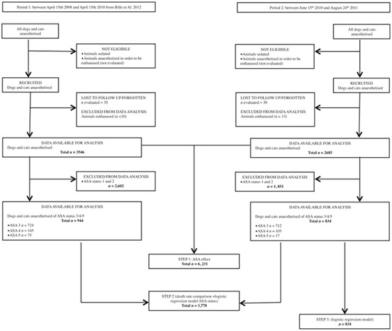
Fig. 1 – Flow diagram of the recruitment and follow-up of the anaesthetic cases.

All dogs and cats that underwent general anaesthesia at the Centre Hospitalier Vétérinaire des Cordeliers during period 1 and period 2 were included. General anaesthesia was defined as a druginduced unconsciousness characterized by a controlled and reversible depression of the central nervous system (CNS) sufficient to allow endotracheal intubation if required. Animals that were sedated or anaesthetized in order to be euthanazed were not eligible. Sedation was defined as chemical restraint insufficient to allow endotracheal intubation.

The patients’ inclusion commenced when general anaesthesia was induced by administration of an injectable induction agent, by whatever route [IV or intramuscular (IM)], or isoflurane until the end of anaesthesia. End of anaesthesia was defined as the return of consciousness characterized by the observation of the patient interacting with its surroundings, rectal temperature above 36°C and sternal recumbency. Animals that were euthanazed during anaesthesia for medical reasons were excluded from the study.

For both periods, death occurring during anaesthesia was recorded.

Data collection

For all patients that were included, identification data, species, age, type of procedure, anaesthetic regimen and epidural administration of a combination of morphine and bupivacaine were recorded.
Prior to induction of anaesthesia, on the basis of history and clinical examination, the attending anaesthetist assessed and recorded the patient’s ASA status. The anaesthetist then decided which anaesthetic regimen would be used.

Following completion of period 1, authors considered that not all available relevant data had been taken into account. Therefore, during period 2, patients’ breed, gender, weight and urgency of the procedure were also recorded.

Grouping

Grouping of patients recruited during period 1 has been described elsewhere (Bille et al. 2012). For patients recruited during period 2, some groupings differed from those described in period 1.

In period 1 animals were grouped according to their actual age. However, in period 2 the animals were grouped in order to reflect the age in relation to the expected life-span of their breed. (Moreau et al. 2003; Adams et al. 2010). The animals’ pedigree papers identified breeds. Animals that were older than the median age at death described for its category were arbitrarily classified as ‘geriatric’. For crossbred dogs, median age at death was considered to be median age at death of the overall population (11.25 years). For cats, a median age at death of 12 years old was retained.

The type of procedure the patient underwent was defined as: ‘examination’ (a non-invasive procedure), ‘orthopaedic’ or ‘soft tissue surgery’. No animal died in the ‘orthopaedic’ category; therefore, ‘orthopaedic’ and ‘soft tissue surgery’ categories were merged into a single ‘surgery’ category (any invasive procedure). If an anaesthetized animal underwent more than one procedure, the most invasive one was retained for the study.

Premedication, anaesthetic induction and maintenance agents were recorded. Twenty-two different premedication regimens were identified. They included the use of acepromazine, midazolam, morphine, fentanyl, butorphanol and medetomidine, used either alone or in combination. For the data analysis, all premedication regimens were gathered in a ‘Yes’ category. The nature of the substance used and the route of administration were not taken into account. Any patient that did not receive premedication was included in a ‘No’ category.

Thirteen different induction regimens were identified. They were grouped into two categories: ‘Propofol’ and ‘alternative induction’. The latter included the use of ketamine, thiopental, fentanyl, isoflurane, medetomidine or dexmedetomidine or its use with ketamine, propofol or thiopental. Two groups of maintenance regimens were defined: patients that received isoflurane and patients that did not.
Patients that did not receive isoflurane were anaesthetized by means of total intravenous anaesthesia or the IM injection of the induction agent with no maintenance.

The anaesthetic regimen was defined by the combination of the premedication, the induction and the maintenance regimens. One hundred and sixteen different anaesthetic regimens were identified. Two main groups were defined:

  • Use of the recommended protocol (premedication, induction of anaesthesia with propofol, maintenance with isoflurane).
  • Use of an alternative anaesthetic regimen. This group included three subgroups:
    • premedication and induction with thiopental, maintenance with isoflurane.
    • premedication and induction with ketamine, maintenance with isoflurane.
    • other (any other combination).

Some variables were only present in period 2. A study by Brodbelt et al. (2008b), found that a bodyweight of <5 kg was a risk factor for anaesthetic-related death in dogs. In period 2 of this current study animals were grouped into three categories: ‘≤5 kg’, ‘>5 kg’ and ‘not weighed’.
The urgency of the procedure was also assessed only in period 2. A procedure was classified as ‘Scheduled’ if staff were notified at least four hours prior to the procedure; as ‘Urgent’ if it was not ‘Scheduled’ but medical management was possible before anaesthesia was induced and as ‘Emergency’ if it was not ‘Scheduled’ and medical management was not possible before anaesthesia was induced.

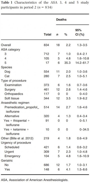
The characteristics of ASA 3, 4 and 5 study participants during period 2 are shown in Table 1. The Centre Hospitalier Vétérinaire des Cordeliers acquired a computed tomography scanner (CT) in December 2009. Details of animals undergoing CT scans were specifically noted.

Bias assessment

To limit inter-observer consistency of the ASA classification the same attending veterinarian (CB) reviewed and, if necessary, reassessed the ASA status on the basis of the patient charts.

The authors aimed to include 950 patients with an ASA status of 3 and over for period 2, in order to reach the same number as in period 1 and allow statistical comparison. However, during the study the sale of the formulation of propofol used in Study 1 was discontinued in France, therefore data collection for the study was terminated on August 24th 2011.

Data analysis

If an animal underwent more than one anaesthesia, each event was considered independently.

The data analysis was conducted following a three-step approach. As a first step, the relationship between death and ASA score was studied. As a second step, because of the very low death rate in animals with an ASA score of 1 or 2 (see Results), the analysis focused on animals with an ASA score of 3 and over. Univariate analysis was performed to compare the characteristic of the two cohorts (Chi square and Mann–Whitney/Wilcoxon Two-Sample Test, EpiInfo 3.5.3; Centers for Disease Control and Prevention, GA, USA). Finally, as a third step, as more variables were available for period 2 than for period 1, a specific analysis was performed on patients with an ASA status of 3 and over in period 2. Univariate analyses were carried out, followed by a multivariate analysis of the data. The statistical unit was the animal and death the outcome variable.

Logistic regression models were created using the GLM procedure of R software 2.13.1 (R Foundation for Statistical Computing, Austria). ‘Period’, ‘ASA category’, ‘species’, ‘type of procedure’, ‘anaesthetic regimen’, ‘urgency of procedure’ and ‘geriatric’ were set as explanatory variables. The probability of wrongly rejecting the null hypothesis was set to 5%. A manual backward procedure was used.

Variables were kept in the model if p < 0.16 and therefore tested for collinearity performing chisquares. Two-way interactions were tested. For assessing the fit of the models, the le Cessie-van Houwelingen-Copas-Hosmer global goodness of fit test were calculated, receiver-operating characteristic (ROC) curves were constructed and the areas under curves (AUCs) were calculated (R software, MKmisc, PresenceAbsence and RelativeRisk libraries).

Animals with missing data (n = 60) were excluded (Fig. 1). This included only animals for which age was missing. The physical status of these animals were ASA 1 (n = 26), ASA 2 (n = 23), ASA3 (n = 10) or ASA4 (n = 1). None of them died during anaesthesia.

Results

Participants and descriptive data

The duration of period 2 (14.3 months) was shorterthan that of period 1 (23.8 months). During the study periods, 6231 animals underwent general anaesthesia; 3546 patients were recruited during period 1 and 2685 during period 2. When considering patients with an ASA status of 3 and over, 944 animals were recruited during period 1 and 834 during period 2. Animals lost to follow-up were estimated to be 60 (30 per period). The number of patients euthanized for medical reasons during general anaesthesia was estimated at <10 during period 1 and was recorded as 13 during period 2 (Fig. 1).

Outcome data

During period 1, 48 animals out of 3546 (1.4%, 95% confidence interval (CI) [1.0–1.7]) died. During period 2, 21 animals out of 2685 (0.8%, 95% CI [0.5–1.2%]) died. No animal died in the ASA 1 category. The death rate increased with each grade of ASA status (Tables 1 and 3).

Main data

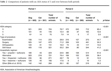
Patients with an ASA status of 3 and over from both periods were compared. Characteristics of the ASA 3, 4 and 5 study participants during both periods are presented in Table 2. There were significantly more cats (p = 0.008) in period 2 (33.6%) than in period 1 (27.6%). ASA status, anaesthetic regimen and type of procedure distributions differed significantly between periods.

During period 1, 45 patients with a status of 3 and over [4.8%, 95% CI (3.5–6.4%)] died under anaesthesia, while during period 2, 18 [2.2%, 95% CI (1.3–3.5%)] died under anaesthesia (Table 2). For these patients, the death rate was significantly lower (p = 0.002) during period 2.

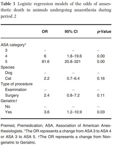
The third-step (multivariate) analysis was performed on the ASA 3, 4 and 5 categories during period 2 (Table 3). No co-linearity was detected between the variables of the model. Dogs had surgery significantly more often than cats but the association between the variables ‘type of procedure’ and ‘Species’ was not strong enough to interfere with the model. A higher ASA status and being geriatric were associated significantly with an increased likelihood of anaesthetic death. Increasing ASA category was very significantly associated with an increased chance of death, and compared to the group ‘Not geriatric’, the group ‘Geriatric’ was associated with a significant 3.6-fold increase in the chance of death. Although not statistically significant, there was a tendency for cats to be more at risk of death during anaesthesia than dogs, and for animals undergoing surgery to be more at risk than those just undergoing examination. After adjusting for other variables, the odds of death associated with patients’ breed, gender, the anaesthetic regimen or urgency of the procedure were not significantly different. The le Cessie-van Houwelingen-Copas-Hosmer global goodness of fit test p value was 0.63. None of the interaction terms were significant and were therefore dropped out of the model. The AUC of the model was 0.80, indicating good discrimination properties (Hosmer & Lemeshow 2000).

Analyses of subgroups and interactions

Characteristics of ‘CT scan’ subgroup are presented in Appendix S2. During period 1, 110 CT scans were performed compared to 453 during period 2. Only those performed on patients of ASA 3, 4 and 5 categories were analyzed. This represented 24 procedures during period 1 and 128 during period 2. Of these, 17 were classified as ‘examination’ in period 1 and 113 in period 2. CT scans represented 6.25% of all ‘examination’ procedures in period 1 and 30.3% in period 2.

Discussion

The overall anaesthetic death rates in period 1 and period 2 were 1.4% and 0.8%, respectively.

As no dog categorized as ASA 1 died, and only one dog as ASA 2 (period 1), subsequent statistical analyses were limited to patients with an ASA status of 3 and over, as will be this discussion unless stated otherwise. In this group, the anaesthetic death rate was significantly lower in period 2. For both periods, results suggest that ASA status was significantly associated with increasing the odds of anaesthetic death in dogs and cats. In period 2, results suggest that ASA status and being geriatric were significantly associated with an increased risk of anaesthetic death in both dogs and cats. There was a tendency for cats and dogs going undergoing surgery (rather than examination) to be more at risk of anaesthetic death. Breed, gender, and urgency of the procedure that took place were not found to be associated with an increased risk of death.

According to EBVM recommendations, the authors used the best available evidence on which to base their recommendations based on the results of period 1 and of other published work. To simplify and increase compliance, only three recommendations were made, chosen by senior staff but, within the limitations below, for all attending veterinarians and nurses (in all, 26 clinical staff members) to take into consideration. To improve ASA status at the time of anaesthesia, pre-anaesthetic medical management of the patient was maximized whenever possible. The recommended anaesthetic regimen for sick patients (premedication, propofol and isoflurane) was based on the following facts: 1) that that premedication appears to decrease the odds of anaesthetic death (Clarke & Hall 1990; Dyson et al. 1998; Brodbelt et al. 2006, 2008a); 2) that the results from period 1 suggested that the use of an anaesthetic regimen including premedication, intravenous thiopental and maintenance with isoflurane increased the risk (Bille et al. 2012), and 3) that results from period 1 also suggested that epidural administration of a combination of morphine and bupivacaine increased risk, possibly through lack of experienced staff administering and monitoring some of these patients.

The attending veterinary surgeon chose the drugs and doses of premedication, and doses of anaesthetic induction agent (sufficient to allow endotracheal intubation) according to the patients needs. In period 2, within the recommendations, veterinarians could include any premedication, induction and maintenance molecules that were available during period 1. For example, low-dose ketamine was routinely used as a perioperative adjunct analgesic.
This access remained unchanged during both periods. If, in a particular situation, the attending veterinarian did not feel comfortable with the recommendations, his or her experience should prevail. As a consequence, senior staff recommendations were not applied in all cases.

When authors compared both periods, significant differences were identified in duration of study period, ASA status, type of procedure, species and anaesthetic regimen (Table 2). Anaesthetic regimen was obviously influenced by the recommendations, and indeed this could introduce a bias in that more very sick animals would receive the regimen ‘premedication-propofol-isoflurane’. It is possible that the lower % of ASA 4 and 5 in period 2 was the result of improved pre-anaesthetic clinical treatment. The increased % of the procedure ‘examination’ probably related to the acquisition of the CT scanner towards the end of period 1, as this increased the number of ‘examinations’ only, for example by contraindicating a surgery that would otherwise have taken place (e.g. identification of metastases). No analytical prospective procedure was performed to evaluate if difference between the periods were caused, in part or totally, by the three senior staff recommendations but it is the authors opinion that they may have contributed, although other factors, not monitored in this study, may also have had an impact.

When comparing both periods, the Centre Hospitalier Vétérinaire des Cordeliers had an increase in overall work load during period 2. During period 1, an average of 150 anaesthetics per month were performed. Of all patients that were presented for consultation, 8.9% were anaesthetized (data not shown). During period 2, an average of 194 anaesthetics per month were recorded. Of all patients that were presented for consultation, 9.3% were anaesthetized (data not shown). This increase in overall workload could, in part, explain why, during period 2, 2685 animals were recruited in only 14.3 months when it took 23.8 months to recruit 3546 patients during period 1. However, it is unlikely that the acquisition of the CT scanner was responsible totally for the difference in durations of study periods.

There was a statistically significant difference (p < 0.001) in ASA status distribution of the participants between both periods. As shown in Table 2, the number of animals classified in ASA 3 category increased by 8.7% in period 2, while the number of animals classified in ASA 4 and 5 categories decreased respectively by 2.8% and 5.9% when compared to period 1. The ASA status was correlated to anaesthetic death during both periods, making it essential to identify reasons explaining the increased recruitment of ASA 3 patients. Judgement of ASA status is a possible source of error. Inter-observer judgment of ASA status has been described as only ‘fair’ in humans (Owens et al. 1978; Burgoyne et al. 2007) and in compromised small animal patients McMillan & Brearley (2013) found major discrepancies between observers. However, we tried to eliminate this discrepancy in our study by having a single investigator re-assess categorization from the records.

The change between the two periods in relationship between ASA status and anaesthetic death was unexpected. As overall death rate decreased in the later period, it could have been expected that death rate would have decreased in the same fashion in each ASA category, but the death rate for the ASA 5 category in period 2 was a much higher proportion than for period 1. In the authors’ opinion, preanaesthetic medical management cannot entirely explain the reduced death rate during period 2.

However, patients still classified as ASA-5 after such intensive medical treatment had demonstrated their inability to respond and would be expected to be at higher risk.

The relationship between ASA categories and the type of procedure performed during both periods was also analyzed. The proportions of animals classified as ASA 3 was higher in period 2, regardless of the type of procedure. During period 1, 110 CT scans were performed compared to 453 during period 2.
Twenty-four CT scans were performed on ASA 3 patients in period 1, compared to 119 in period 2 (Appendix S2). The acquisition of a CT scanner might explain the increased recruitment of ASA 3 patients during period 2. The acquisition of a CT scanner undoubtedly modified ASA distribution of the patients. It does account for part of the increased recruitment of the ‘ASA 3’ ‘examination’ category.

However, it does not explain the higher proportion of animals classified as ASA 3 and going through surgery, nor the overall decrease in ASA 4 and 5 patients.

Cats were significantly more represented (p = 0.008) in period 2 (33.6%) than in period 1 (27.6%) (Table 2). The type of procedure cats underwent was statistically significantly different (p = 0.0006) (data not shown) between the two periods. When compared to dogs, the proportions of cats classified in the ‘examination’ and ‘soft tissue’ categories were similar in both periods. The proportion of cats classified in the ‘orthopaedics’ category was 16% higher in period 2. In cats, none of the CT scans performed (n = 28) were followed by surgery (data not shown). They were all classified in the ‘examination’ category (data not shown). The reason why the recruitment of cats during period 2 increased remains unclear. Interestingly, a larger number of cats went through orthopaedic procedures in period 2. These patients had not undergone a prior CT scan. Recruitment of cats was not in parallel with the recruitment for of dogs for CT scan examination.

The use of the recommended anaesthetic regimen in ASA-3 patients (premedication, -propofol -isoflurane) represented 23.6% of the 944 anaesthetics in period 1 and 61.6% of the 834 regimens in period 2 (Table 2) demonstrating that senior staff recommendations were understood and well applied by the staff. The absence of an association between the anaesthetic regimen used and death occurring during anaesthesia was unexpected; As the senior staff recommended that animals with an ASA status of 3 and over should receive an anaesthetic regimen including premedication, induction with propofol and maintenance with isoflurane, it was expected that this group would have a higher death rate than the ‘alternative regimen’ group. However, the potential recruitment bias might have been overestimated, and as previously stated, if the attending veterinarian did not feel comfortable with recommendations, his or her experience could prevail
Also, authors underestimated the proportion of cats whose aggressive behaviour prevented staff from gaining IV access, and therefore where anaesthetic induction was with IM injection or isoflurane inhalation. These animals were included in the ‘alternative regimen’ group. In both periods, about 50% of the cats fell into the ‘other regimen’ group (Table 2).

Finally, one dog died during induction of anaesthesia and was included in the ‘alternative regimen’ group because it died before receiving isoflurane. If it had survived, it would have been classified in the ‘premedication + propofol + isoflurane’ group.

There are a number of limitations to this study. No pre-study power calculation was performed to to quantify the level of odds ratios likely to be able to detect for the risk factors, but in fact period 2 was terminated when propofol was not available. The control group was historical rather than concurrent, but a random study would not have been practicable.
The decrease in the occurrence of anaesthetic death may implies a significant improvement in patient care over the study periods, but this improved care may not only be due to the recommendations, but to improved practice protocols in general, including surgical practice. Another factor possibly influencing the trend to lower death rates include an increase in the number of staff, attending veterinarians and nurses. Such limitation to assessing the results has been described in human literature (Keenan & Boyan 1991; Sprung et al. 2003; An et al. 2011). The variables that surround peri-anaesthetic patient management and their complex interactions are numerous. If senior staff recommendations had a beneficial effect on death rate, they can only account for part of the modifications that were described.

A further limitation of this study is that data recorded during both periods could not be analyzed statistically in the same fashion. The choice of anaesthetic regimen was influenced in period 2, creating a selection bias. Also, the number of epidural analgesics in period 2 were too low to be analyzed. The new variables thought to be of clinical importance (e.g. breed, gender, urgency of the procedure and old age) could not be compared between both periods. In period 1, animals were classified according to their chronological age, and no statistical differences were found in risk. In period 2 animals were classified in relation to the longevity of the breed- for example, a 10 year-old Great Dane should not be considered the same way as a 10-yearold Yorkshire terrier. With classification, the risk of death during anaesthesia in geriatric animals was shown to be increased.

Finally, some grouping could not be done in the same fashion during both periods; as explained earlier, in period 2 no animal died in the ‘orthopaedic’ category so ‘orthopaedic’ and ‘soft tissue surgery’ categories were merged into a ‘surgery’ category (any invasive procedure). These limitations had consequences on the data analysis. Originally, it was intended to generate a logistic regression model including both periods. Such a model was generated (data not shown), and included ASA status, species and period as variables. Interactions between ASA status and period were statistically significant. Due to its statistical complexity, this model carried a high risk of misinterpretation, and it is not presented here.
Instead, a logistic regression model was generated with different variables, only for period 2 (step 3, Table 3).

It is of major importance to emphasize that senior staff recommendations were defined after analysis of their pertinence and applicability in our facility. EBVM principles state that best available evidence should be integrated into the facility’s clinical expertise, and the drugs available. New drugs (e.g. alfaxalone) may play a part, and another team, in another clinical setting, may therefore well come to different recommendations.

In conclusion, the results of this study support the hypothesis that anaesthesia safety improved in our practice between the two study periods. Application of evidence-based veterinary medicine to anaesthetic death in small animal practice lead to the formulation of three recommendations for the clinical staff at the practice, which may have played a role in decreasing the anaesthetic death rate. Other factors, not monitored in this study, may also have had an impact.

References

  1. Adams VJ, Evans KM, Sampson J et al. (2010) Methods and mortality results of a health survey of purebred dogs in the UK. J Small Anim Pract 51, 512–524.
  2. Alef M, Von Praun F, Oechtering G (2008) Is routine preanaesthetic haematological and biochemical screening justified in dogs? Vet Anaesth Analg 35, 132–140.
  3. An J, Li-ming Z, Sullivan Erin A et al. (2011) Intraoperative cardiac arrest during anesthesia: a retrospective study of 218 274 anesthetics undergoing non-cardiac surgery in a US teaching hospital. Chin Med J 124, 227–232.
  4. Bille C, Auvigne V, Libermann S et al. (2012) Risk of anaesthetic mortality in dogs and cats: an observational cohort study of 3546 cases. Vet Anaesth Analg. 1, 59–68.
  5. Brodbelt DC, Hammond R, Tuminaro D et al. (2006) Risk factors for anaesthetic-related death in referred dogs. Vet Rec 158, 563–564.
  6. Brodbelt DC, Pfeiffer DU, Young LE et al. (2007) Risk factors for anaesthetic-related death in cats: results from the confidential enquiry into perioperative small animal fatalities (CEPSAF). Br J Anaesth 99, 617–623.
  7. Brodbelt DC, Blissitt KJ, Hammond RA et al. (2008a) The risk of death: the confidential enquiry into perioperative small animal fatalities. Vet Anaesth Analg 35, 365– 373.
  8. Brodbelt DC, Pfeiffer DU, Young LE et al. (2008b) Results of the confidential enquiry into perioperative small animal fatalities regarding risk factors for anesthetic-related death in dogs. J Am Vet Med Assoc 233, 1096–1104.
  9. Burgoyne LL, Smeltzer MP, Pereiras LA et al. (2007) How well do pediatric anesthesiologists agree when assigning ASA physical status classifications to their patients? Paediatr Anaesth 17, 956–962.
  10. Clarke KW, Hall LW (1990) A survey of anaesthesia in small animal practice: AVA/BSAVA report. Vet Anaesth Analg 17, 4–10.
  11. Dodman NH, Lamb LA (1992) Survey of small animal anesthetic practice in Vermont. J Am Anim Hosp Assoc 28, 439–444.
  12. Dyson DH, Maxie MG, Schnurr D (1998) Morbidity and mortality associated with anesthetic management in small animal veterinary practice in Ontario. J Am Anim Hosp Assoc 34, 325–335.
  13. Gaynor JS, Dunlop CI, Wagner AE et al. (1999) Complications and mortality associated with anesthesia in dogs and cats. J Am Anim Hosp Assoc 35, 13–17.
  14. Gil L, Redondo JI (2013) Canine anaesthetic death in Spain: a multicentre prospective cohort study of 2012 cases. Vet Anaesth Analg 2013, e57–e67.
  15. Hosgood G, Scholl DT (1998) Evalution of age as a risk factor for perianesthetic morbidity and mortality in the dog. J Vet Emerg Crit Care 8, 222–236.
  16. Hosgood G, Scholl DT (2002) Evaluation of age and American Society of Anesthesiologists (ASA) physical status as risk factors for perianesthetic morbidity and mortality in the cat. J Vet Emerg Crit Care 12, 9–15.
  17. Hosmer DW, Lemeshow S (2000) Applied Logistic Regression, Wiley Series in Probability and Mathematical Statistics, October 2000 (2nd edn). John Wiley, New York, USA. pp. 392. ISBN: 978-0-471-35632-5.
  18. Joubert KE (2000) Routine veterinary anaesthetic management practices in South Africa. J S Afr Vet Assoc 71, 166–172.
  19. Keenan RL, Boyan CP (1991) Decreasing frequency of anesthetic cardiac arrests. J Clin Anesth 3, 354–357.
  20. McMillan M, Brearley J (2013) Assessment of the variation in American Society of Anesthesiologists [corrected] Physical Status Classification assignment in small animal anaesthesia. Vet Anaesth Analg 40, 229–236.
  21. Moreau D, Cathelain P, Lacheretz A (2003) Comparative study of causes of death and life expectancy in carnivorous pets (II). Revue Med Vet 154, 127–132.
  22. Owens WD, Felts JA, Spitznagel EL (1978) ASA physical status classifications: a study of consistency of ratings. Anesthesiology 49, 239–243.
  23. Redondo JI, Rubio M, Soler G et al. (2007) Normal values and incidence of cardiorespiratory complications in dogs during general anaesthesia. A review of 1281 cases. J Vet Med A 54, 470–477.
  24. Robertson SR (2007) Refining the clinical question: the first step in evidence-based veterinary medicine. Vet Clin North Am Small Anim Pract 37, 419–431.
  25. Sackett DL, Rosenberg WM, Gray JA et al. (1996) Evidence based medicine: what it is and what it isn’t. BMJ 312,71–72.
  26. Schmidt PL (2007) Evidence-based veterinary medicine: evolution, revolution, or repackaging of veterinary practice? Vet Clin North Am Small Anim Pract 37, 409–417.
  27. Sprung J, Warner ME, Contreras MG et al. (2003) Predictors of survival following cardiac arrest in patients undergoing noncardiac surgery: a study of 518,294 patients at a tertiary referral center. Anesthesiology 99, 259.
  28. Williams LS, Levy JK, Robertson SA et al. (2002) Use of the anesthetic combination of tiletamine, zolazepam, ketamine, and xylazine for neutering feral cats. J Am Vet Med Assoc 220, 1491–1495.

Received 7 June 2012; accepted 21 January 2013.

Supporting Information

Additional Supporting Information may be found in the online version of this article:

  • Appendix S1. Logistic regression modelling of the odds of anaesthetic death in animals undergoing anaesthesia during period 1 (Bille et al. 2012).
  • Appendix S2. Characteristics of the patients who went through a CT Scanner, for both periods (n = 563).
Retour

Informations pratiques

CHV des Cordeliers

Retour
  • Lun
    8h - 12h 13h30 - 19h
  • Mar
    8h - 12h 13h30 - 19h
  • Merc
    8h - 12h 13h30 - 19h
  • Jeu
    8h - 12h 13h30 - 19h
  • Vend
    8h - 12h 13h30 - 19h
  • Sam
    8h - 12h 13h30 - 17h30
  • Dim
    Urgences 24h/7j
Retour

Adresse de la clinique

29 - 35 Av. du Maréchal Joffre 77100 MEAUX
retrouvez-nous sur Google Maps