Page d'accueil de Cordeliers
  • Accueil et urgences01 64 34 11 55

Atrial cardiomyopathy

in an adult Labrador retriever dog

A 7-year-old castrated male Labrador retriever was examined for a 10-day history of weakness and syncope. Physical examination revealed bradycardia and a grade III/VI left apical systolic heart murmur. Electrocardiography demonstrated bradycardia, absence of P waves and an atrio-ventricular nodal escape rhythm.


Auteurs : Drs. E. Bomassi1, J.-F. Rousselot2, S. Libermann1, I. Raymond Letron3, S. Etchepareborde1, C. Misbach1  16-01-2017
E-mail : ebomassi@chvcordeliers.com
Cet article a été publié dans : Band 159, Heft 11, November 2017 p 4-6©


1Centre Hospitalier Vétérinaire des Cordeliers, 29-35 avenue du Maréchal Joffre, 77100 Meaux.
2Clinique Vétérinaire des Camélias, Colombes, France.
3INP-ENVT, Université de Toulouse, Laboratoire d’Histopathologie Expérimentale et Comparée et STROMALab UMR5273 UPS EFS INSERM U1031, Toulouse, France

Atrial cardiomyopathy in an adult Labrador retriever dog

Abstract

A 7-year-old castrated male Labrador retriever was examined for a 10-day history of weakness and syncope.
Physical examination revealed bradycardia and a grade III/VI left apical systolic heart murmur. Electrocardiography demonstrated bradycardia, absence of P waves and an atrio-ventricular nodal escape rhythm. Echocardiography revealed marked biatrial enlargement. Thoracic radiographs showed no evidence of pulmonary edema.
Routine plasma biochemistry and electrolytes, basal serum cortisol, total thyroxin concentration, and complete blood count were within normal limits. Serum cardiac troponin I concentration was moderately increased. Serological examinations for antibodies against vector-borne diseases were negative.
A pacemaker was implanted one month after the initial presentation due to worsening of the dog’s clinical condition despite medical treatment. The dog remained asymptomatic for 18 months but was then re-presented with a gastric dilatation volvulus and subsequently euthanized.
Necropsy and histology of the heart yielded a diagnosis of atrial cardiomyopathy.

Atriale Kardiomyopathy bei einem erwachsenen Labrador Retriever

Ein 7-jähriger, kastrierter Labrador wurde für Leistungsschwäche während 10 Tagen und Synkope vorgestellt. Die Auskultation ergab eine Bradykardie und ein Herzgeräusch (Grad III/VI) über dem linken Herzapex. Das EKG zeigte eine Bradykardie, keine P Wellen und einen junktionalen (AV) Ersatz-Rhythmus. Über eine Echokardiographie konnte eine signifikante, bi-atriale Kammer-Vergrößerung dargestellt werden, aber auf dem Thorax-Röntgenbild wurde kein Lungenödem festgestellt. Blutanalysen ergaben normale Werte sowohl für Blutchemie, Serumelektrolyte, basales Serumkortisol, totales Thyroxin und Differenzialblutbild. Das kardiale Serum-Troponin war mittelmäßig erhöht. Die Antikörper-Serologie für Vektor-übertragene Krankheiten war negativ. Einen Monat nach der ersten Präsentation wurde ein Herzschrittmacher implantiert, da die klinischen
Symptome schlimmer wurden, trotz medikamentöser Behandlung. Anschließend blieb der Hund asymptomatisch, aber 18 Monate später wurde er erneut vorgestellt und wegen eines Magen Dilatation-Volvulus euthanasiert. Mittels Autopsie und histologischer Untersuchung wurde eine atriale Kardiomyopathie diagnostiziert. 

Case history

A 7-year-old 35 kg, neutered male Labrador retriever dog was examined for a 10-day history of weakness and syncope (5-6 per day). On physical examination, the dog was in good clinical condition. Cardiac auscultation revealed bradycardia (heart rate: 50 beats/min) and a grade III/VI left apical systolic heart murmur. The rest of the physical examination was unremarkable.

Electrocardiography

Standard 6-lead electrocardiography (ECG) showed bradycardia, absence of P wave and a regular (ie, RR interval variation less than 10%) atrio-ventricular nodal escape rhythm at a rate of 58 beats/minute (bpm, Fig. 1).

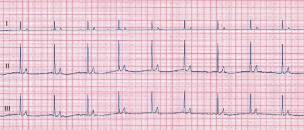
Figure 1 – Standard 6-lead electrocardiogram tracing obtained at 25 mm/s and 10 mm/mV. Note the absence of consistently definable P waves and the narrow-complex junctional escape rhythm with a rate of 58 bpm.

Echocardiography

Conventional echocardiography and standard Doppler examination were performed on the awake dog in standing position with an ultrasound unit (Megas and My Lab Twice, Esaote Biomedica, Firenze, Italy).

The left ventricular M-mode echocardiogram obtained from the right parasternal transventricular short-axis view revealed a moderate left ventricular dilation in diastole (51.0 mm) with normal systolic diameter (26.5 mm) (reference ranges (RR) of 34.0-46.3 mm and 20.5-30.8 mm, respectively, Gonçalvez et al., 2002).

The fractional shortening was moderately increased (48%, RR = 29-43%, Chetboul et al., 2005).

The two70 dimensional right parasternal transaortic short-axis view showed marked left atrial (LA) enlargement (LA to aorta ratio = 2.52, RR < 1.6, Rishniw and Erb, 2000).

The right atrium appeared dilated on the two-dimensional right parasternal long axis view with a ratio between the right and left atria of 0.8 (Fig. 2).
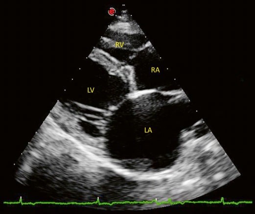
Figure – Extract of the 24-hour Holter examination (leads I and II) obtained at 25 mm/s and 10 mm/mV. Note the absence of consistently definable P waves, the narrow-complex junctional escape rhythm (mean rate of 58 bpm) and the periods of ventricular escape beats.

The aspect of all cardiac valves was within normal limits except for mild thickening of the mitral leaflets.

The left apical 4-chamber view revealed moderate mitral regurgitation using Color Doppler mode (area of the regurgitation jet signal to LA area < 50%).

Finally, pulsed and continuous Doppler mode showed an increased E mitral wave velocity, reflecting LA pressure overload (2.68 m/s, RR: 0.61-1.13 m/s, Chetboul et al., 2005) and the absence of diastolic transmitral atrial phase flow.

Radiography

Thoracic radiographs revealed cardiomegaly (vertebral heart scale of 12.5, RR < 10.7, Buchanan and Bücheler, 1995) and pulmonary venous congestion but no signs of cardiogenic pulmonary edema.

Holter examination

Twenty-four-hour Holter monitoring (Fig. 3) confirmed the presence of persistent bradyarrhythmia (mean heart rate: 52 bpm) associated with ventricular escape beats at lower heart rate.

No episode of syncope was reported during the Holter examination.

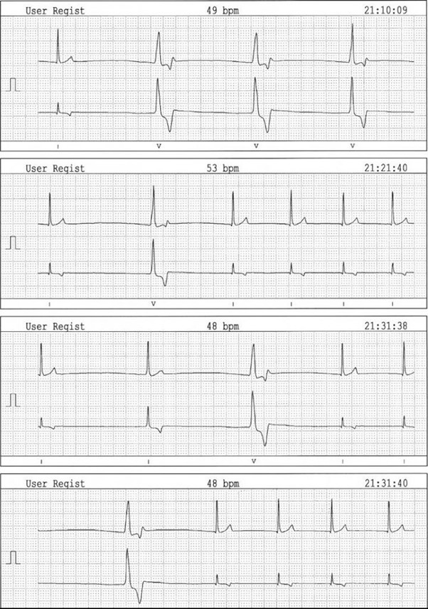
Figure 3 – Image of a two-dimensional right parasternal long axis 4-chamber view. Note the biatrial enlargement and moderate thickening of the mitral valve leaflets. LV: left ventricle, RV: right ventricle, RA: right atrium, LA: left atrium

Blood analysis

Routine plasma biochemistry and electrolytes, serum basal cortisol, total thyroxin concentrations and complete blood count were within normal limits. Serum cardiac troponin I concentration was moderately increased (0.712 ng/mL, RR < 0.06 ng/mL, Laboratoire Idexx, Alfort, France).

Finally, a qualitative ELISA test (SNAP 4Dx Test, Idexx Laboratories, Westbrook, USA) failed to detect any antibodies against vector-borne diseases such as erhlichiosis, anaplasmosis, dirofilariosis and Lyme disease.

Therapy and follow-up

The dog was medicated per os with benazepril (Fortekor, Novartis Santé Animale, Rueil Malmaison, France), 0.28 mg/kg SID, furosemide (Dimazon, Intervet, Beaucouze, France), 0.57 mg/kg BID, prednisolone (Megasolone, Merial, Lyon, France), 0.57 mg/kg BID and doxycycline (Ronaxan, Merial, Lyon, France), 8.6 mg/kg SID.

Due to the increase of syncopal episodes (ie, > 10 per day), a pacemaker implantation was scheduled one month after the initial presentation. A single chamber, permanent pacemaker (Adapta ADSR01, Medtronic, Minneapolis, USA) with VVIR (ventricular pacing, ventricular sensing, inhibition response and rate-adaptive; lower heart rate: 70 bpm; upper tracking rate: 130 bpm; amplitude: 3,5 V; pulse width : 0,64 ms) combined with a bipolar epicardial wire (CapSure Epi, Medtronics Boulogne-Billancourt, France) was implanted using a standard surgical approach (Monnet 2003).

Several left ventricular myocardial biopsies were taken during the procedure. Histopathological examination failed to reveal any degenerative, inflammatory or infectious disease of the ventricular myocardial and epicardial tissues.

Prednisolone and doxycycline were discontinued 10 days after surgery. Follow-up included clinical, ECG, Holter and echocardiographic examinations.

Despite a decrease in serum cardiac troponin I concentration (0.105 versus 0.712 ng/mL) and ventricular diameter (48.5 versus 51.0 mm) 4 weeks after pacemaker implantation, the atria remained dilated with an LA to aorta ratio of 2.19 (versus 2.52 at first examination).

The dog remained asymptomatic and in good clinical condition for 18 months but was then presented with gastric dilatation volvulus resulting in euthanasia.

Gross examination revealed the atria to be highly enlarged and discolored with thin walls. The ventricles were moderately dilated and the atrio-ventricular valve leaflets were thickened and irregular.

Histological examination of both atria revealed severe extensive replacement fibrosis and atrophic modifications of the residual myofibers that were dissociated by interstitial collagen deposition (Fig. 4). Foci of mild interstitial fibrosis were found in the ventricles.

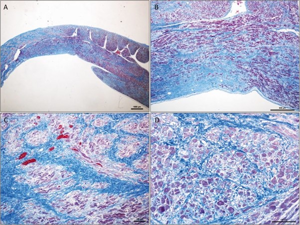
Figure 4 – Microscopic aspect of the right atrial wall at low magnification. Severe extensive interstitial fibrosis dissociating
and replacing the myocardial fibers. Residual myofibers are dissociated by interstitial collagen deposition and exhibit atrophic changes. Masson trichrome stain, original magnification ×10 (A), ×40 (B), ×100 (C), ×200 (D)

Discussion

The present report describes an exclusive atrial cardiomyopathy (AC) characterized by fibrous infiltration of the atrial myocardium and atrophy of atrial cardiomyocytes in an adult Labrador retriever dog.

The AC was associated with a bradyarrhythmia causing weakness and syncope, which are frequent presenting complaints in dogs with bradyarrhythmia (Wess et al., 2006).

Similar AC have been previously described in dogs, including one male crossbred Chow134 chow (Une et al.,1998), 2 mongrels and 4 Springer Spaniels (Buchanan, 2005), two Labrador retrievers (one female and one male, Schmitt and Lefbom, 2016) and one Greyhound (Wesselowski et al., 2017). Most of these dogs had clinical signs compatible with congestive heart failure and bradyarrhythmia, such as abdominal distention and syncope.

In all cases, necropsy and histopathological examination revealed enlarged, thin-walled and pale atria, fibrous infiltration of the atrial myocardium (with or without fatty degeneration) and atrophy and loss of the atrial cardiomyocytes (Une et al., 1998; Buchanan, 2005; Schmitt and Lefbom, 2016; Wesselowski et al., 2017).

Some dogs showed different degrees of chronic inflammation of the atrial myocardium, associated in a few dogs with concomitant atrial and ventricular involvement (Buchanan, 2005).

Unlike the dog in the present report, most of these dogs were young, i.e., between 12 and 36 months old, and one dog was 4.5 years (versus 7 years in the present case).

This suggests that AC may have different etiologies, as observed in humans (Goette et al, 2016). Interestingly, 8 of the 10 dogs were diagnosed with atrial standstill, confirmed by ECG (Une et al., 1998; Buchanan, 2005; Wesselowski et al., 2017).

Thus, this arrhythmia seems to be frequently associated with AC, as already highlighted in several other reports in which AC was suspected (MacAulay, 2002; Nakamura et al., 2012; Serene, 2012; Thomason et al., 2016; Cervenec et al., 2017).

In the present case, ECG examination revealed an absence of definable P waves, a nearly regular junctional escape rhythm and ventricular escape beats. Differentials included therefore atrial standstill, sinus arrest and atrial fibrillation with complete atrio-ventricular block that might have been differentiated by carrying out an electrophysiologic study (not performed here for practical reasons).

In the present case, and in agreement with previous reports (Schmitt and Lefbom, 2016; Wesselowski et al., 2017), echocardiography was relatively non-specific of AC and showed marked biatrial enlargement, mild ventricular diastolic dilation (which could be attributed to bradycardia) with nearly normal fractional shortening and moderate mitral valve regurgitation.

The latter was probably secondary to both mitral annulus dilation and degeneration of the mitral valve leaflets because this LA enlargement could not be explained by the magnitude of regurgitation alone. Moreover, the moderate increase of serum troponin I concentration reflected mild myocardial injury and excluded active myocarditis.

Nevertheless, the evolution of previous subclinical atrial myocarditis into chronic AC could not be excluded with certainty. Finally, pacemaker implantation led to complete resolution of symptoms and a survival time of 18 months. During follow-up, the decrease in serum troponin I concentration and left ventricular diameter was attributed to the regression of bradyarrhythmia. However, the atria remained greatly enlarged with an LA to aorta ratio of 2.19 (versus 2.52 at first examination). In the literature, 2 dogs with AC and bradyarrhythmia were successfully managed for approximately 7 years after artificial pacing.

The main complications included progressive worsening of atrial dilation, left ventricular dilation with decreased ventricular contractility, and development of congestive heart failure 3 to 4 years after pacemaker implantation.

According to the EHRA/HRS/APHRS/ SOLAECE expert consensus group in human medicine (Goette et al., 2016), AC is defined as “Any complex of structural, architectural, contractile or electrophysiological changes affecting the atria with the potential to produce clinically-relevant manifestations”. In humans, AC may be primary (e.g., isolated atrial amyloïdosis, ‘lone’ atrial fibrillation, hereditary muscular dystrophies, mutation of the gene encoding for the precursor protein for atrial natriuretic peptide) or secondary to various cardiac and extra-cardiac diseases such as congestive heart failure, cardiac valvulopathies, myocarditis, diabetes mellitus, obesity and ageing. In the present case, no underlying cause was identified and the AC was considered idiopathic. This report has several limitations.

Firstly, skeletal muscle biopsies could have been done to explore muscular dystrophy butwere not envisaged in view of the absence of clinical signs related to muscular disease.Moreover, an electrophysiological study would have been interesting to characterize thebradyarrhythmia as well as advanced echocardiographic techniques such as speckle trackingimaging to evaluate atrial function.

In conclusion, the present case is the third AC confirmedby histology in a Labrador retriever dog. As previously reported, pacemaker implantationseems to be effective for managing bradycardia-associated syncope in this uncommon heartdisease. Further studies are needed to better understand AC and possibly identify a breedpredisposition in Labrador retriever dogs, as already suggested in English Springer Spaniels.

Acknowledgments

The authors are sincerely grateful to Dr Anna Gelzer for the English to German translation.

Reference

  1. Buchanan J. W., Bücheler J.: Vertebral scale system to measure canine heart size in radiographs. J. Am. Vet. Med. Assoc. 1995, 206: 194–199.
  2. James Buchanan Cardiology Library [database on the Internet] Davis, California: Veterinary Information Network, Inc c2000. Available from http://www.vin.com/library/general/JB106atrialStandstill.htm Last accessed 20 June, 2016.
  3. Cervenec R. M., Stauthammer C. D., Fine D. M., Kellihan H. B., Scansen B. A.: Survival time with pacemaker implantation for dogs diagnosed with persistent atrial standstill. J. Vet. Cardiol. 2017 (in press).
  4. Chetboul V., Sampedrano Carlos C., Concordet D., Tissier R., Lamour T., Ginesta J., Gouni V., Nicolle A. P., Pouchelon J. L., Lefebvre H. P.: Use of quantitative two-dimensional color tissue Doppler imaging for assessment of left ventricular radial and longitudinal myocardial velocities in dogs. Am. J. Vet. Res. 2005, 66: 953–961.
  5. Gonçalves A. C., Orton E. C., Boon J. A., Salman M. D.: Linear, logarithmic, and polynomial models of M-mode echocardiographic measurements in dogs. Am. J. Vet. Res. 2002, 63: 994–999.
  6. Goette A., Kalman J. M., Aguinaga L., Akar J., Cabrera J. A., Chen S. A., Chugh S. S., Corradi D., D’Avila A., Dobrev D., Fenelon G., Gonzalez M., Hatem S. N., Helm R., Hindricks G., Ho S. Y., Hoit B., Jalife J., Kim Y. H., Lip G. Y., Ma C. S., Marcus G. M., Murray K., Nogami A., Sanders P., Uribe W., Van Wagoner D. R., Nattel S.: EHRA/HRS/APHRS/SOLAECE expert consensus on Atrial cardiomyopathies: Definition, characterisation, and clinical implication. J. Arrhythm. 2016, 32: 247–278.
  7. MacAulay K.: Permanent transvenous pacemaker implantation in an Ibizan hound cross with persistent atrial standstill. Can. Vet. J. 2002, 43: 789–791.
  8. Monnet E.: Pacemaker Therapy. In: Ed. D. Slatter, Textbook of small animal surgery, Elsevier Science, 2003, 3rd ed., pp 932–943.
  9. Nakamura R. K., Russell N. J., Shelton G. D.: Adult-onset nemaline myopathy in a dog presenting with persistent atrial standstill and primary hypothyroidism. J. Small. Anim. Pract. 2012, 53: 357–360.
  10. Rishniw M. and Erb H. N.: Evaluation of four 2-dimensional echocardiographic methods of assessing left atrial size in dogs. J. Vet. Intern. Med. 2000, 14: 429–435.
  11. Schmitt K. E., Lefbom B. K.: Long-term management of atrial myopathy in two dogs with single chamber permanent transvenous pacemakers. J. Vet. Cardiol. 2016, 18: 187–193.
  12. Serene R. L.: Atrioventricular muscular dystrophy in a 5-month-old English springer spaniel. Can. Vet. J. 2009, 50: 1286–1287.
  13. Thomason J. D., Kraus M. S., Fallaw T. L., Calvert C. A.: Survival of 4 dogs with persistent atrial standstill treated by pacemaker implantation. Can. Vet. J. 2016, 57: 297–298.
  14. Une Y., Furusawa N., Shirota K., Nomura Y.: Atrial adipofibrosis in a dog. J. Vet. Diagn. Invest. 1998, 10: 192–194.
  15. Wess G., Thomas W. P., Berger D. M., Kittleson M. D.: Applications, complications, and outcomes of transvenous pacemaker implantation in 105 dogs (1997-2002). J. Vet. Intern. Med. 2006, 20: 877–884.
  16. Wesselowski S., Abbott J., Borgarelli M., Tursi M.: Presumptive partial atrial standstill secondary to atrial cardiomyopathy in a Greyhound. J. Vet. Cardiol. 2017 (in press).
Retour

Informations pratiques

CHV des Cordeliers

Retour
  • Lun
    8h - 12h 13h30 - 19h
  • Mar
    8h - 12h 13h30 - 19h
  • Merc
    8h - 12h 13h30 - 19h
  • Jeu
    8h - 12h 13h30 - 19h
  • Vend
    8h - 12h 13h30 - 19h
  • Sam
    8h - 12h 13h30 - 17h30
  • Dim
    Urgences 24h/7j
Retour

Adresse de la clinique

29 - 35 Av. du Maréchal Joffre 77100 MEAUX
retrouvez-nous sur Google Maps