Page d'accueil de Cordeliers
  • Accueil et urgences01 64 34 11 55

Left Ventricular Propagation

Sommaire

  • Echocardiography is currently an obligate tool when studying cardiac diseases and their consequences in domestic carnivores.
  • This test allows the identification of anatomic, morphologic (valvular, myocardial, large vessels) and hemodynamic (blood flows) abnormalities seen with cardiac diseases.

Auteur : Dr. E. Bomassi 05-07-2012 Centre Hospitalier Vétérinaire des Cordeliers, 29 avenue du Maréchal Joffre, 77100 Meaux. E-mail : ebomassi@chvcordeliers.com

Clinical application of the left ventricular flow propagation velocity with Color M-mode Doppler in 26 dogs with mitral valve disease

Echocardiography is currently an obligate tool when studying cardiac diseases and their consequences in domestic carnivores. This test allows the identification of anatomic, morphologic (valvular, myocardial, large vessels) and hemodynamic (blood flows) abnormalities seen with cardiac diseases. Several modes of data acquisition are available for this purpose: 2D and M-mode imaging, conventional Doppler and more recently tissue Doppler Imaging (tDI) 1-3.

Logical assessment of some abnormalities gives an idea of myocardial performance and functioning. Ventricular function is composed of a diastolic phase and a systolic phase. Echocardiographic measurement indices (either measured or calculated) are already available for each phase taken separately or together. These indices are diverse and can be obtained with the echocardiographic modes described previously 4, 5. Transmitral flow with a pulsed-wave Doppler mode is most commonly used in general practice for evaluation of diastolic function 6-8.

Transmitral flow is composed of an early-diastole E wave (passive ventricular filling), and an end-diastole A wave (active ventricular filling, associated with atrial contraction). In the normal dog the mitral E/A ratio varies between 1.04 and 2.42 9.

Three main abnormal flow profiles are recognized 10-12: type 1 profile, where ventricular relaxation is impaired when the filling pressures during end-diastole are reduced (“abnormal relaxation”); type 2 profile, also called restrictive where ventricular compliance is impaired and filling pressures are very high during end-diastole (“restrictive”); type 3 profile, where the relaxation is impaired and filling pressures are moderately high during end-diastole (“pseudonormal”). Even though these mitral indices are easy to obtain and therefore appealing, their use is however limited because they combine ventricular relaxation and loading conditions in evaluating diastolic function 13, 14. Moreover, these indices can vary, depending on the heart rate 13, 14.

It is therefore sometimes difficult to estimate the diagnostic value of some abnormalities because their origin can be multifactorial, for example when a type 3 flow (“pseudo-normal”) is identical to a normal flow 13-15.

Other indices for evaluation of diastolic function have been studied in domestic carnivores. Left ventricular flow propagation velocity (Vp) can be obtained by pulsed-wave Color M-mode Doppler imaging and is a new index of diastolic function 16 that has been experimentally validated in healthy dogs and cats 17, 18.

Unlike with transmitral flows, this velocity does not depend on loading conditions and heart rate 19, 20, but only on ventricular relaxation19, 20. When combined with mitral E wave velocity (pulse-wave Doppler), the E/Vp ratio can be obtained which does not depend on ventricular relaxation, but only on loading conditions 21, 22.

Measurement of Vp allows the establishment of an independent index of ventricular relaxation, and its ratio with the E wave provides an independent index which reflects end diastole filling pressures. This measure has been experimentally validated in the normal dog. The purpose of this study was to investigate the use of Vp and E/Vp as indices of evaluation of diastolic function and filling pressures in the dog with MVD.

Our first hypothesis was that Vp would be significatively different between healthy dogs and dogs with relaxation abnormalities, when comparing the different Appleton flows, allowing a distinction between a normal profile (healthy dog) and a type 3 “pseudonormal” profile. Our second hypothesis was that it is possible to use the ratio E/Vp for the evaluation of filling pressures during end-diastole, in contrast to Appleton profiles.

figure 1

Materials and methods

The animals were presented to the Cardiology Service by their owners. One group of dogs was healthy, with no morphologic or hemodynamic cardiac abnormalities; the other groups of dogs suffered from acquired MVD with mitral insufficiency which had not yet been treated at that time. The diagnosis of cardiac failure was made based on clinical signs according to ISACHC classification criteria 23, or if thoracic radiographs showed evidence of congestive heart failure.

The dogs were divided in four groups:

  • group 1: normal dogs (control group) (normal stage) ;
  • group 2: dogs with MVD and a type 1 mitral profile (abnormal relaxation) (stage 1) ;
  • group 3: dogs with MVD and a type 3 mitral profile (pseudonormal) (stage 3) ;
  • group 4: dogs with MVD and a type 2 mitral profile (restrictive filling) (stage 2) ;

Dogs with a disease other than cardiac in origin, or with a concomitant disease were not included in the study. Dogs with a cardiac disease other than MVD, or dogs MVD undergoing treatment (that could influence and alter echocardiographic results) and dogs presented on an emergency basis were excluded from the study.

The Tukey test was performed between groups to test for differences regarding age, weight, heart rate (HR) and shortening fraction (SF). A linear correlation was performed to test relation between age, weight, HR and SF and Vp and E/Vp. For each parameter (E, A, Vp and E/Vp), 5 successive measures were taken on 5 consecutive cardiac cycles.

Mean and standard deviation were calculated. For each parameter (E, A, Vp and E/Vp), mean, standard deviation, median, maximum and minimum values were calculated for each group. For each group, Vp and E/Vp distribution were compared with non parametric tests because the small number of animals in each group was not representative of a normal distribution.

Therefore an ANOVA on ranks procedure using the Dunn test was performed to test for differences between groups. A value of p < 0.05 (2-sided) was considered significant. Results are expressed in mean (median) ± standard deviation.

The transmitral valve flow profile was obtained with left parasternal imaging using an apical four-chamber long axis view with pulsed-wave conventional Doppler. The sample volume was positioned just below the mitral annulus. E wave and A wave were measured in m/s.

The different mitral types (1, 2 and 3) corresponded to those described before 10-12 :

  • type 1: small E wave, large A wave, increase in the deceleration time of E wave, ratio E/A < 1 ;
  • type 2: large E wave, small A wave, decreased deceleration time, ratio E/A increased (> 2.42) 9 ;
  • type 3: E and A waves of normal magnitude, normal deceleration time, ratio E/A within normal limits (between 1.04 and 2.42) 9.

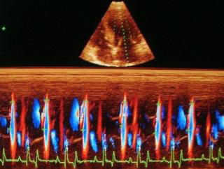
The left ventricular flow propagation velocity (Vp) was obtained with a Color M-mode pulsed-waved Doppler with a left apical long axis image, four chamber view. From this image, and with two-dimensional echocardiography, the color box was positioned in order to sample the entire surface of the left ventricle, from the mitral annulus to the apex 20.

Aliasing was programmed to be between 50 % and 75 % of the maximal velocity of the E wave of the transmitral flow 20. The M-mode cursor was then positioned in the continuity of the ventricular color flow during diastole. The M mode was then activated with a maximum sweep speed (50 mm/s).

Vp was measured as the slope during the first aliasing of E wave from the mitral annulus to the apex of the left ventricle. It was measured in m/s. For each parameter (E, A, Vp, ratio E/Vp), 5 successive measures were taken on 5 consecutive cardiac cycles. Mean and standard deviation were then calculated.

Résults

  • 26 dogs were included in the study:
    • group 1: normal dogs (control group): 5 dogs ;
    • group 2: dogs with MVD and abnormal relaxation mitral profile: 7 dogs ;
    • group 3: dogs with MVD and pseudonormal mitral profile: 8 dogs ;
    • group 4: dogs with mitral MVD and restrictive mitral profile: 6 dogs.
  • There were 12 females and 14 males. The mean age was 8.5 ± 3.3 years.
  • The age of the dogs in group 4 was significantly higher (p < 0.05) than group 1.
  • There were no differences between the other groups regarding age and no differences between all groups regarding weight.
  • Dogs of group 4 attended to have higher ISACHC class than the other groups.
tableau 1

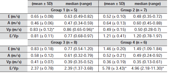
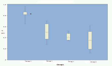
There was a significant difference for Vp between group 1 (normal dogs) and groups 2-4 (dogs with MVD) (p < 0.05). There was no difference between the different groups of dogs with MVD (group 2-4). There was no significant correlation between age, weight, HR or SF and Vp.

  
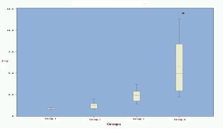
There was a significant difference for E/Vp between group 4 (restrictive profile) and group 1 (normal dogs), group 2 (abnormal relaxation), and group 3 (pseudonormal) (p < 0.05). There was no difference between groups 1, 2 and 3. There is no significant correlation between age, weight, HR or SF and E/Vp.

Discussion

The results confirms our first hypothesis that Vp can be used to distinguish between healthy and sick dogs, regardless of the stage of diastolic dysfunction.

Indeed, there was a significant difference between healthy and sick dogs, within the different groups, and globally. This is of particular interest in distinguishing between a normal and “pseudo-normal” mitral profile, because the spectral aspect of E and A waves, their maximum velocity, and their ratio are identical in these cases. However, Vp does not allow the distinction between the different groups of sick dogs, because the values are not statistically different between the different Appleton flows.

Vp is a load-independent index of ventricular relaxation that has been experimentally and clinically validated in man 16, 17, 19-22, and thus experimentally validated in dogs and cats 17, 18. Based on the results of this study we propose that it can be considered as an index of left ventricular relaxation in clinical practice in the dog.

As far as the E/Vp is concerned, the results confirm our second hypothesis on the role of this ratio. The theory of the E/Vp ratio is to get an independent calculated index which reflects filling pressure. Indeed, E wave velocity mainly depends on relaxation and preload 10-12. Vp only depends on relaxation, and is perfectly correlated with tau 17. E/Vp ratio therefore only depends on the preload.

The results indicate that E/Vp allows the distinction between sick dogs of group 4 (stage 2 Appleton with restrictive flow and with the most elevated flow propagation pressures) and all other dogs. This allows the distinction between a restrictive profile and all other profiles (normal, abnormal relaxation and pseudonormal). Unlike Vp for relaxation, E/Vp allows the evaluation of high degrees of filling pressures depending on the Appleton stages. On the other hand, there is no difference between normal dogs and dogs with a type 1 and 3 profile.

This can be explained by the absence or low increase in filling pressures in the latter ones. In man, normal E/Vp ratio is less than 1.5. There is an intermediate zone between 1.5 and 2.5 which is difficult to interpret. Abnormal E/Vp is more than 2.5 28, 30, 33. It is considered that a ratio of less than 2 indicates pressures less than 15 mmHg, and a ratio of greater than 2.5 indicates elevated pressures above 15 mmHg 34, 35, with a sensitivity of 79 % and a specificity of 77 % 36, 37.

In man a validated formula to calculate the pulmonary capillary pressure 21 : Pcp = 5.27 x (E/Vp) + 4.6 (in mmHg) is used. These data are consistent with our results in the dog, because in the normal dog E/Vp is normal, in the sick dogs at stage 1 E/Vp is still normal, in stage 3 E/Vp is border line and in stage 2 E/Vp is elevated. As for Vp, the objective of this study was not to establish reference values for this ratio; however, our results are similar to what it observed in man. Thus the formula for calculation of pulmonary capillary pressures could potentially also be applied in dogs, although further validation in the dog is required.

The main limitation of this study is the small number of animals, especially for Vp. Another limitation of this study is the lack of evaluation of influence of mitral regurgitation on the propagation velocity. Severe mitral regurgitation increases E wave velocity of mitral inflow due to the volume overload.

Theorically Vp should’nt be influenced by volume overload. The last limitation of this study is the other lack of evaluation of influence of age on E, A an Vp in normal dogs. As reported in healthy man 38 negative correlation exists for E and Vp with age. In our study, there is no correlation between age and Vp (linear correlation), but normal and abnormal dogs are considered. Further studies including a larger number of dogs should be performed in the future.

Bibliographie

  1. Chetboul V et coll (2004) Quantification, repeatability, and reproducibility of feline radial and longitudinal left ventricular velocities by tissue Doppler imaging. Am J Vet Res 65: 566-572.
  2. Chetboul V et coll (2004) Use of tissue Doppler imaging to confirm the diagnosis of dilated cardiomyopathy in a dog with equivocal echocardiographic findings. J Am Vet Med Assoc 225: 1877-1880.
  3. Chetboul V et coll (2004) Tissue Doppler assessment of diastolic and systolic alterations of radial and longitudinal left ventricular motions in Golden Retrievers during the preclinical phase of cardiomyopathy associated with muscular dystrophy. Am J Vet Res 65: 1335-1341.
  4. Boon JA (1998) The echocardiographic examination. In: Manual of veterinary echocardiography (Boon JA, Edr) The Williams & Wilkins Co, Baltimore.
  5. Lee BH et coll (2002) Evaluation of a novel Doppler index of combined systolic and diastolic performance in Newfoundland dogs with familial prevalence of dilated cardiomyopathy. Vet Radiol Ultrasound 43: 154-165.
  6. Schober KE et coll (1998) Pulmonary venous flow characteristics as assessed by transthoracic pulsed Doppler echocardiography in normal dogs. Vet Radiol Ultrasound 39: 33-41.
  7. Luis Fuentes V (2003) Diastolic function – is this the key to successful management of many feline cardiomyopathies? J Feline Med Surg 5: 51-56.
  8. Schober K, Luis Fuentes V (1998) Doppler echocardiographic assessment of left ventricular diastolic function in dogs. Tierärztl Prax Ausg Klientiere Heimtiere 26: 13-20.
  9. Boon JA (1998) Echocardiographic reference values. In: Manual of veterinary echocardiography (Boon JA, Edr) The Williams & Wilkins Co, Baltimore, 453-473.
  10. Appleton CP et coll (1988) Relation of transmitral velocity patterns to left ventricular diastolic function: new insights from a combined hemodynamic and Doppler study. J Am Coll Cardiol 12: 426-440.
  11. Appleton CP, Hatle LK (1992) The natural history of left ventricular filling abnormalities: assessment by two-dimensional and Doppler echocardiography. Echocardiography 9: 437-457.
  12. Klein AL et coll (1990) Serial Doppler echocardiographic follow-up of the left ventricular diastolic
  13. Rossi A et coll (2001) Mitral regurgitation and left ventricular diastolic dysfunction similarly affect mitral and pulmonary vein flow Doppler parameters: the advantage of end-diastolic markers. J Am Soc Echocard 14: 562-568.
  14. Rossvoll O, Hatle LK (1993) Pulmonary venous flow velocities recorded by transthoracic Doppler ultrasound: relation to left ventricular diastolic pressures. J Am Coll Cardiol 21: 1687-1696.
  15. Zhang H et coll (2002) Noninvasive differentiation of normal from pseudonormal/restrictive mitral flow using TEI index combining systolic and diastolic function. Circulation 66: 831-836.
  16. Garcia MJ et coll (1998) New Doppler Echocardiographic Applications for the Study of Diastolic Function. J Am Coll Cardiol 32: 865-875.
  17. Garcia MJ et coll (2000) Color M-mode Doppler flow propagation velocity is a preload insensitive index of left ventricular relaxation: animal and human validation. J Am Coll Cardiol 35: 200-208.
  18. Schober KE et coll (2003) Comparison between invasive hemodynamic measurements and noninvasive assessment of left ventricular diastolic function by use of Doppler echocardiography in healthy anesthetized cats. Am J Vet Res 64: 93-103.
  19. Garcia MJ et coll (1999) Color M-mode Doppler flow propagation velocity is a relatively preload-independent index of left ventricular filling. J Am Soc Echocard 12: 129-137.
  20. Brun P et coll (1992) Left ventricular flow propagation during early filling is related to wall relaxation: a color M-mode Doppler analysis. J Am Coll Cardiol 20: 420-432.
  21. Garcia MJ et coll (1997) An index of early left ventricular filling that combined with pulsed Doppler peak E velocity may estimate capillary wedge pressure. J Am Coll Cardiol 29: 448-454.
  22. Stugaard M et coll (1994) Abnormalities of left ventricular filling in patients with coronary artery disease: assessment by color M mode Doppler technique. Eur Heart J 15: 318-327.
  23. Miller MS, Tilley LP (1995) The International Small Animal Cardiac Health Council (ISACHC). Recommendations for the diagnosis and the treatment of heart failure in small animals In Manual of Canine and Feline Cardiology. WB Saunders, Philadelphia, 469-502.
  24. Chetboul V et coll (2005) Effects of animal position and number of repeated measurements on selected two-dimensional and M mode echocardiographic variables in healthy dogs. J Am Vet Med Assoc 227: 743-747.
  25. Oyama MA, Sisson DD (2005) Assessment of cardiac chamber size using anatomic M-mode. Vet Radiol Ultrasound 46: 331-336.
  26. Schober KE, Maerz I (2005) Doppler echocardiographic assessment of left atrial appendage flow velocities in normal cats. J Vet Cardiol 7: 15-25.
  27. Dukes McEwan J et coll (2002) Doppler echocardiography in the dog : measurement variability and reproductibility. Vet Radiol Ultrasound 43: 144- 153.
  28. Drighil A et coll (2002) Study of variations in preload on the new echocardiographic parameters of diastolic function in the healthy subject. Arch Mal Cœur 95: 573-580.
  29. Nishihara K et coll (2000) Usefulness of early diastolic flow propagation velocity measured by color M-mode Doppler technique for the assessment of the left ventricular diastolic function in patients with hypertrophic cardiomyopathy. J Am Soc Echocard 13: 801-808.
  30. Donal E et coll (2002) Paramètres doppler dans le suivi et l’évaluation des patients insuffisants cardiaques. Arch Mal Cœur Vaiss 95: 123-128.
  31. Dougherty AH et coll (1984) Congestive heart failure with normal systolic function. Am J Cardiol 54: 778-782.
  32. Litwin SE, Grossman W (1993) Diastolic dysfunction as a cause of heart failure. J Am Coll Cardiol 22: 49-55.
  33. Abergel E, Habib G (2003) Bilan hémodynamique et données pronostiques obtenues par échographie chez l’insuffisant cardiaque. Arch Mal Cœur Vaiss 96: 43-50.
  34. Abergel E (2005) Indices échocardiographiques de fonction diastolique à l’épreuve de la pratique. Arch Mal Cœur Vaiss 98: 325-330.
  35. Gonzalez-Vilchez F et coll (1999) Combined use of pulsed and color M-mode Doppler echocardiography for the estimation of pulmonary capillary wedge pressure: an empirical approach based on an analytical relation. J Am Coll Cardiol 34: 515-523.
  36. Nagueh SF et coll (1997) Doppler tissue imaging: a noninvasive technique for evaluation of left ventricular relaxation and estimation of filling pressures. J Am Coll Cardiol 30: 1527-1533.
  37. Ommen SR (2001) Echocardiographic assessment of diastolic function. Curr Opin Cardiol 6: 240-245.
  38. Mego DM et coll (1998) Variation of flow propagation velocity with age. J Am Soc Echocardiogr 11: 20-25.
Retour

Informations pratiques

CHV des Cordeliers

Retour
  • Lun
    8h - 12h 13h30 - 19h
  • Mar
    8h - 12h 13h30 - 19h
  • Merc
    8h - 12h 13h30 - 19h
  • Jeu
    8h - 12h 13h30 - 19h
  • Vend
    8h - 12h 13h30 - 19h
  • Sam
    8h - 12h 13h30 - 17h30
  • Dim
    Urgences 24h/7j
Retour

Adresse de la clinique

29 - 35 Av. du Maréchal Joffre 77100 MEAUX
retrouvez-nous sur Google Maps