Page d'accueil de Cordeliers
  • Accueil et urgences01 64 34 11 55

ERG findings

in three hypothyroid adult dogs with and without levothyroxine treatment

Abstract

Purpose To evaluate the effects of levothyroxine (LTh) on the electroretinogram (ERG) of adult dogs.

Material and methods Binocular, full field photopic and scotopic ERGs were recorded from an anesthetized Maltese Bichon cross (MB), a Yorkshire Terrier (YT) and a Shetland Sheepdog (SS) affected with hypothyroidism and treated with a daily dose of LTh at 20μg/kg. The photopic ERGs were evoked to 12 different intensities ranging from 0.81 to–2.19 log cd.s/mand presented under photopic conditions in order to assess (from the derived luminance-response curves) Vmax and b : a amplitude ratio parameters. Photopic flicker ERGs were obtained at 30 Hz. The scotopic ERGs (intensity: –3.09 log cd.s/m2) were recorded while the retina was dark-adapting and after 32 min of dark adaptation.


Auteurs : Drs. Philippe Durieux,* F. Rigaudière,† J.-F. LeGargasson‡ and S. G. Rosolen
*Clinique Vétérinaire, Meaux, France;†UDD Paris 7, France; INSERM U-592, UMR UPMC-Paris 6, Institut de la Vision, Paris, France; CliniqueVétérinaire, Asnières, France; Fondation Ophtalmologique A. de Rothshild, Paris, France

Centre Hospitalier Vétérinaire des Cordeliers, 29 avenue du Maréchal Joffre, 77100 Meaux.
Mots clefs/Key Words : dog, electroretinogram, hypothyroidism, levothyroxine, photopic ERG, scotopic ERG
E-mail : pdurieux@chvcordeliers.com
Cet article a été publié dans : Veterinary Ophthalmology (2008) 11, 406–411

This procedure was performed on two separate sessions: following a 3-day interruption of LTh treatment (S1) and following 30 days without interruption of LTh treatment (S2).

Results
The mean photopic a-wave peak times were 9.8 ms at S1 and 5.0 ms at S2, respectively. The mean photopic b-wave peak times were 23.3 ms at S1 and 11.5 ms at S2, respectively, and the mean scotopic b-wave peak times (after 32 min of dark adaptation) were 45.2 ms at S1 and 26.0 ms at S2, respectively. No other significant ERG changes were observed.

Conclusion
Our results indicate that a dose of 20 μg/kg of LTh given to adult dogs was accompanied by a marked peak time shortening of both photopic and scotopic ERGs, without affecting other ERG parameters.

Introduction

Hypothyroidism is one of the most common endocrinopathy diagnosed in adult dogs.1 While it is not the purpose of this paper to review clinical signs, tests for diagnosis, the treatment with synthetic levothyroxine (LTh)2 is both straightforward and effective3 with a continuous decrease of thyroid volume.4 Because of the increase in medical care of dogs and the efficacy of the treatment, a long-term administration of LTh is often performed.

It is well known that the fundamental action of thyroid hormone (TH) is exerted on protein metabolism to regulate multiple metabolic processes.5 The TH is essential for the normal development of the central nervous system (CNS). 6,7 Along with other components of the developing CNS the neural retina is a known target of TH signaling. 8 Within the retina, photoreceptor development is particularly sensitive to manipulations of TH signaling in many species such as fish 9 and other vertebrates. 10,11 Recently, many studies have demonstrated the implication of TH in cone differentiation 12 during the retinal development, growth and regeneration. 13,14 Few findings are documented in adult dogs that have been receiving LTh treatment for many months and sometimes many years.

Relatively little attention has been given to the effect of TH on the mature nervous and sensory systems. The electroretinogram (ERG), a commonly used technique for evaluating the photopic and scotopic retinal function, has been used for many years in veterinary ophthalmology. 15,16

The purpose of this study was to evaluate, with the ERG, if the retinal function of dogs affected with hypothyroidism and treated with daily oral administration of LTh is altered.

Materials and methods

Animals

The following inclusion criteria were used:

  • hypothyroidism diagnosed for at least 2 years based on positive biological and clinical signs (i.e. alopecia, squamosis, hyperseborrhea, bradycardia, obesity, fatigability, and hypercholesterolemia), total T4 lower than 16 nm/L (CERI, Paris, France), basal cTSH > 0.8 ng/mL (CERI),
  • complete remission of biological and clinical signs of hypothyroidism following treatment with levothyroxine (Levothyrox®, Merck-Lipha-Santé, Lyon, France; 20μg/kg per day).

Based on these criteria, three dogs were included in this study, namely, a male Bichon Maltese Bichon cross (MB;age: 10 years old; weight: 3.5 kg; cTSH at time of diagnosis: 1.5 ng/mL), a male Yorkshire Terrier (YT; age: 13 years old; weight: 3.5 kg; cTSH at time of diagnosis: 1.3 ng/mL) and a male Shetland Sheepdog (SS; age: 9 years old; weight: 15 kg; cTSH at time of diagnosis: 2.1 ng/mL).

All dogs were found to be in good physical condition following a complete clinical examination to determine if they could be safely anesthetized. A complete ophthalmological examination, including slit-lamp and indirect opththalmoscopy performed 72 h before each ERG session, did not reveal signs of an underlying retinopathy. A blood sample was performed prior to anesthesia in order to measure T4 before each ERG recording session.

Prior to anesthesia, the pupils were maximally dilated with 1% tropicamide (Mydriaticum®, Théa, France) to obtain a stable mydriasis at least 1 h before the recording session. Pupil diameter was measured before and after the ERG examination with the aid of a strabismus compass to ascertain that maximal pupillary dilation had been achieved and maintained.

The dogs were anesthetized with a single intramuscular injection of a mixture of ketamine (5 mg/kg, Imalgène®, Mérial, Lyon, France) and medetomidine (0.1 mg/kg, Domitor®, Pfizer, Orsay, France). A hot water blanket was used to maintain the body temperature of anesthetized dogs. The dogs were intubated to maintain the airway and allow for ventilation if required. The entire procedure lasted 45 min approximately.

Material

A Visiosystem (Siem Bio-Médicale, Nîmes, France) was used to generate the light stimuli (single flash and flicker) as well as record and analyze the ERG responses. Binocular ERGs were evoked with the use of two bright flashes of light (Xenon capacitive discharge adjustable photostimulators; maximum intensity: 0.81 log cd.s/m2; duration: 20μs) delivered in full field condition as previously reported.17

The eyes were maintained opened and the third eyelid prolapse avoided with the use of sterile sclero-conjunctival copper clips attached to the conjunctiva of the superior part of the globe. 18 These clips also served as the active ERG electrodes. Reference and ground electrodes were inserted subcutaneously at the level of the outer canthus and neck, respectively (sterile acupuncture needles of 0.2 mm in diameter manufactured by Asiamed, Paris, France). Corneal hydration was maintained throughout the entire procedure with the use of a carbopol gel (Ocrygel®, TVM, Paris, France).  The ERG responses were amplified 10 000 times within a 0.1–300 Hz recording bandwidth (6 db per octave) and recorded over a 250-m period for flash ERGs and 500 ms for flicker ERGs.

ERG procedure

The retinal signals were obtained in photopic and scotopic conditions as previously described. 17,18 Briefly, following a preadaptation period of 3 h to a photopic background (acclimatization
period), the animals were prepared as described above and the cone function was tested against a photopic (rod-desensitizing) background of 30 cd/m2 measured at the level of the dog’s eyes (Digital Lux tester YF-1065, Paris, France). The photopic (cone-mediated) responses were evaluated first. Photopic single flash ERGs were evoked in response to 12 different flashes of decreasing intensities (maximum intensity: 0.81 log cd.s/m2, minimum intensity: –2.19 log cd.s/m2 ; average of 15 flashes at interstimulus intervals of 1.3 Hz). These data were then used to derived the photopic luminance-response function curve, as well as the ERG b-wave/a-wave amplitude ratio.

The Naka–Rushton equation [V=Vmax I(n)/(I(n) + K(n)] was used to calculate Vmax. 18,19 Vmax is the asymptotic value of the b-wave amplitude as a function of stimulus luminance, K is the intensity that produces a b-wave whose amplitude is half of Vmax and n is a dimensionless constant that controls the slope of the function and represents the degree of homogeneity of retinal sensitivity.

The photopic flicker ERG responses evoked to flashes of Imax in intensity (e.g. flash intensity that yields the Vmax) derived from the luminance response curve were evoked at a temporal frequency of 30 Hz delivered for 20 s.

Following the assessment of the cone-mediated function, the rod desensitizing background lights were then turned off and averages of five flashes of dim blue light [Kodak Wratten filter 98 (440 nm) intensity: –3.09 log cd.s/m2] delivered at a temporal frequency of 0.1 Hz were obtained at the onset of the dark adaptation process (t0) and, thereafter, at regular time intervals (t0t4t8t16 and t32 min) during dark adaptation.

The above protocol was repeated in two different occasions: Session 1, immediately following a 3-day interruption of LTh treatment and Session 2, following 30 days without any interruption of LTh treatment. In the latter case, the ERG examination always took place 7 h after LTh intake.

Data analysis

The amplitude of the ERG a-wave was measured from the baseline to a-wave trough and that of the b-wave, from trough of the a-wave to the peak of the b-wave. Similarly, peak times were measured from flash onset to the peak of individual waves. For the photopic flicker ERG, amplitudes were measured from baseline to peak.

RESULTS

Pupil diameter remained constant throughout the entire recording session. Similarly, there were only minor changes in rectal temperature.

Individual T4 serum concentration values are presented in Table 1a. For all animals tested in Session 1, the T4 serum concentration, measured before each ERG session, were below the normal range (Table 1). Similarly, in Session 2, all animals yielded T4 values that were within the range of the usual values given by the referring laboratory (Table 1).

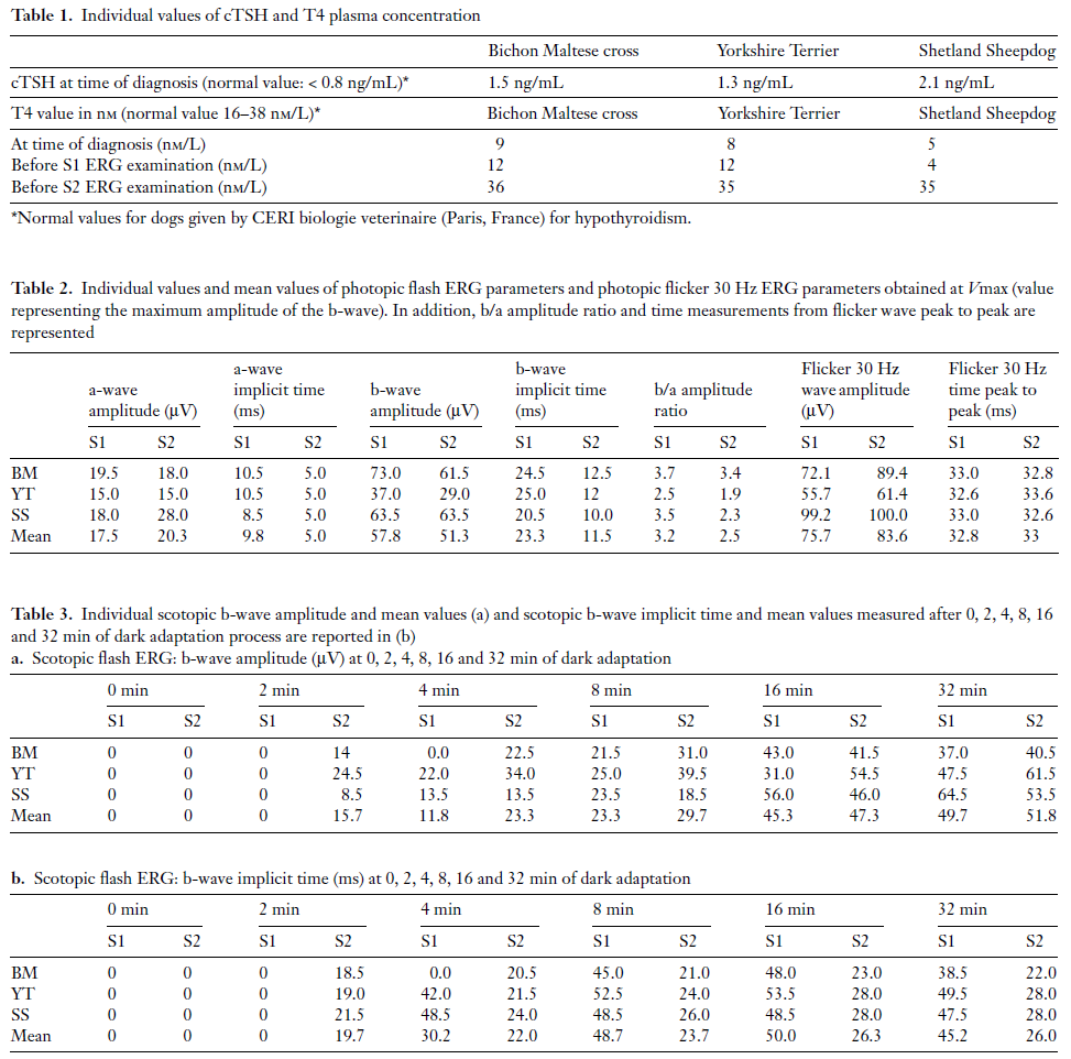
Photopic and scotopic ERG parameters measured for each of the three dogs are reported in Tables 2 and 3, respectively, while representative ERG responses are shown in Fig. 1. Amplitude measurements (a-wave, b-wave amplitudes, flicker, b/a ratio and log k) of Sessions 1 and 2 were not different from each other. In contrast, a- and b-wave implicit times were shorter in Session 2 compared to Session 1.

Figure 1. Representative ERGs (right eye, RE; left eye, LE) of the Shetland Sheepdog (SS)
obtained in photopic conditions: flash at Vmax (AS1 and AS2) and flicker 30 Hz (BS1 and BS2),
and in scotopic conditions at 32 min of dark adaptation (CS1 and CS2). The vertical bar (0) represents the flash onset.

Discussion

Our results indicate that the dose of 20 μg/kg of LTh given once daily induced, as expected, a rapid and significant increase in T4 serum concentration. This increase in T4 serum concentration was accompanied by a marked shortening (nearly half of normal value) of the peak times of the photopic a- and b-waves and scotopic b-wave while the flicker response was not affected.

It is true that we did not have access to baseline (before the hypothyroidism) ERG measures from the three dogs and therefore two scenarios are possible, namely, normal ERG in S2 and delayed in S1 (hypothyroidism effect) or normal ERG in S1 and faster in S2 (drug effect). Of interest, a brief survey of the literature 20–22 indicates that the peak times of the cone ERG in dogs (irrespective of breed) will vary between 11 and 14 ms for the a-wave and 25–34 ms for the b-wave compared to 47–67 ms for the rod b-wave. These values are well within the range of our S1 ERG measures, suggesting that LTh intake accelerated the retinal responses. Following thyroidectomy in adult rat, Takeda et al23 showed that this surgical procedure caused a lengthening of the peak time of the ERG b-wave indicating that the retina is highly sensitive to TH. Recently, it has been demonstrated that TH is required to maintain the rate of renewal of the outer segments of the photoreceptor cell. 24 Similarly, patients with idiopathic hypothyroidism were shown to present with an abnormal increase in implicit times and subnormal amplitude of ERG responses before treatment. 25 After treatment with thyroxine the patient became euthyroid and repeat ERGs disclosed normal retinal function.

According to our results, it would appear that the effect of LTh intake in hypothyroidism dogs accelerates the implicit time of the photopic and scotopic ERG responses. Although our results are different from those previously reported in rats and humans, they nonetheless clearly suggest a drug effect on the dog retina, especially at the level of photoreceptors. This hypothesized echanism of action at the level of the photoreceptor is also supported by the absence of significant alteration of the ERG flicker response which is said to result from the interaction of the ON and OFF retinal pathways 26 at the postreceptoral level. 27

Although our dogs suffered from hypothyroidism, they had been treated for at least 2 years prior to this study and given that the treatment was stopped only 3 days prior to ERG recording, it could be that the effect of hypothyroidism on the retina was too short to yield a significant result. This could also explain the difference between our study and that reporting rat and human results.

We realize that our report is only based on three dogs and despite rarity of this condition we should try to increase the number of observations in order to achieve statistical significance. However, despite this cautionary note, we believe that one should take into consideration LTh intake when conducting a retinal assessment in hypothyroid dogs, given that a positive sign of treatment success will markedly impact on the timing of the ERG.

Acknowledgments

We are grateful for helpful comments from Prof. Pierre Lachapelle (Department of Ophthalmology, McGill University Montreal Children Hospital, Quebec, Canada) and Dr Dan Rosenberg (Ecole Nationale Véterinaire d’Alfort, Maisons-Alfort, France), internist.

References

1. Fergusson DC. Testing for hypothyroidism in dogs. Veterinary Clinics of North American Small Animal Practice 2007; 37 : 647–669.
2. Panciera DL. Hypothyroidism in dogs: 66 cases (1987–92). Journal of American Veterinary Medical Association 1994; 204 : 761–767.
3. Greco DS, Rosychuk RA, Ogilvie GK et al. The effect of levothyroxine treatment on resting energy expenditure of hyptothyroid dogs. Journal of Veterinary Internal Medicine 1998; 12: 7–10.
4. Taeymans O, Daminet S, Duchateau L et al. Pre- and post-treatment ultrasonography in hypothyroid dogs. Veterinary Radiology and Ultrasound 2007; 48: 262–269.
5. Zhang J, Lazar MA. The mechanism of action of thyroid hormones. Annual Revue of Physiology 2000; 62: 439–466.
6. Dussault JH, Ruel J. Thyroid hormones and brain development. Annual Revue of Physiology 1987; 49: 321–334.
7. Sagham JY. Thyroid disease: an overview. Radiology and Technology 2001; 73: 25–40.
8. Harpavat S, Cepko CL. Thyroid hormone and retinal development: an emerging field. Thyroid 2003; 13: 1013–1019.
9. Browman H, Hawryshyn C. Retinoic acid modulates retinal development in the juveniles of a teleost fish. Journal of Experimental Biology 1994; 193: 191–207.
10. Kelley MW, Turner JK, Reh TA. Ligands of steroid/thyroid receptors induce cone phtotreceptors in vertebrate retina. Development 1995; 121: 3777–3785.
11. Flamant F, Samarut J. Thyroid hormone receptors: lessons from knockout and knock-in mutant mice. Trends in Endocrinology Metabolism 2003; 14: 85–90.
12. Ng L, Hurley JB, Dierks B et al. A thyroid hormone receptor that is required for the development of green cone photoreceptors. Nature Genetics 2001; 27: 94–98.
13. Mader MM, Cameron DA. Photoreceptor differentiation during retinal development, growth, and regeneration in a metamorphic vertebrate. Journal of Neuroscience 2004; 24: 11463–11472.
14. Mader MM, Cameron DA. Effects of induced systemic hypothyroidism upon the retina: regulation of thyroid hormone receptor alpha and photoreceptor production. Molecular Vision 2006; 12: 915–930.
15. Ofri R. Clinical electrophysiology in veterinary ophthalmology: the past, present and future. Documenta Ophthalmologica 2002; 104: 5–16.
16. Narfstrom K, Ekesten B, Rosolen SG et al. Guidelines for clinical electroretinography in the dog. Documenta Ophthalmologica 2002; 105: 83–92.
17. Rosolen SG, Rigaudière F, Lachapelle P. A practical method to obtain reproducible binocular electroretinogram in dogs. Documenta Ophthalmologica 2002; 105: 93–103.
18. Rosolen SG, Rigaudière F, Le Gargasson J-F et al. Recommendations for a toxicological screening ERG procedure in laboratory animals. Documenta Ophthalmologica 2005; 110: 57–66.
19. Rufiange M, Rousseau S, Dembinska O et al. Cone-dominated ERG luminance-response function: the Photopic Hill revisited. Documenta Ophthalmologica 2002; 104: 231–248.
20. Maehara S, Osawa A, Itoh N et al. Detection of cone dysfunction induced by dogoxin in dogs by multicolour electroretinography. Veterinary Ophthalmology 2005; 8: 407–413.
21. Maehara S, Itoh N, Wakaiki S et al. The effects of cataract stage, lens-induced uveitis and cataract removal on ERG in dogs with cataract. Veterinary Ophthalmology 2007; 10: 308–312.
22. Ropstad EO, Bjerkas E, Narfstrom K. Electroretinographic findings in the Standard Wire Haired Dachshund with inherited early onset cone-rod dystrophy. Documenta Ophthalmologica 2007; 114: 27–36.
23. Takeda M, Onoda N, Suzuki M. Characterization of thyroid hormone effect on the visual system of the adult rat. Thyroid 1994; 4: 467–474.
24. Takeda M, Kakegawa T, Suzuki M. Effect of thyroidectomy on photoreceptor cells in adult rat retina. Life Science 1996; 58: 631–637.
25. Holder GE, Condon JR. Pattern visual evoked potentials and pattern electroretinograms in hypothyroidism. Documenta Ophthalmologica 1989; 73: 127–131.
26. Bush RA, Sieving PA. Inner retinal contributions to the primate photopic fast flicker electroretinogram. Journal of the Optical Society of American Association 1996; 13: 557–565.
27. Kondo M, Sieving PA. Primate photopic sine-wave flicker ERG: vector modelling analysis of component origins using glutamate analogs. Investigative Ophthalmology and Visual Science 2001; 42: 305–312.

© 2008 American College of Veterinary Ophthalmologists

Retour

Informations pratiques

CHV des Cordeliers

Retour
  • Lun
    8h - 12h 13h30 - 19h
  • Mar
    8h - 12h 13h30 - 19h
  • Merc
    8h - 12h 13h30 - 19h
  • Jeu
    8h - 12h 13h30 - 19h
  • Vend
    8h - 12h 13h30 - 19h
  • Sam
    8h - 12h 13h30 - 17h30
  • Dim
    Urgences 24h/7j
Retour

Adresse de la clinique

29 - 35 Av. du Maréchal Joffre 77100 MEAUX
retrouvez-nous sur Google Maps