Page d'accueil de Cordeliers
  • Accueil et urgences01 64 34 11 55

Tumor Necrosis Fatcor alpha

Sommaire

Concentration en TNF-alpha dans l’humeur aqueuse de chiens sains ou atteints d’une pathologie intraoculaire : une étude pilote préliminaire

Summary

Objectives

To determine reference values of tumor necrosis factor-alpha (TNF-α) concentrations in the aqueous humor of control dogs. To show whether these values are significantly
different from those obtained in dogs affected with intraocular pathology: acute anterior uveitis (AAU) or chronic primary angle closure glaucoma (PACG).

Auteurs : Drs. P. Durieuxa,∗, S. Etcheparebordea, D. Fritzb, S.G. Rosolenc, d 27-01-2016

a Centre Hospitalier Vétérinaire des Cordeliers, 29 avenue du Maréchal Joffre, 77100 Meaux.
b Companion animal laboratory (C.A.L), 1, rue Salomon-Rachi, 10000 Troyes, France
c Inserm, U968, UPMC université Paris 06, UMR-S968, institut de la vision, 17, rue Moreau, 75012 Paris, France
d Clinique vétérinaire Voltaire, 19, boulevard Voltaire, 92600 Asnières-sur-Seine, France
E-mail : pdurieux@chvcordeliers.com
Cet article a été publié dans : Journal français d’ophtalmologie (2015) 38, 288—294

Tumor necrosis factor-alpha concentration in the aqueous humor of healthy and diseased dogs: A preliminary pilot study

Methods

Forty-four dogs were included in the study and were divided into two groups: a control group and a group with intraocular disease. Twenty-seven dogs (9 males and 18 females) were examined and found to be normal after a complete ophthalmological examination (control group), 7 (6 females and 1 male) presented with PACG, and 10 (7 females and 3 males) presented with AAU secondary to corneal perforation. One aqueous humor sample (volume ≥ 0.2 mL) was collected from one eye of all dogs. The aqueous TNF-α concentration was determined with an Elisa kit.

Results

TNF-α levels were detectable in all dogs. TNF-α levels were significantly higher in the group with intraocular disease compared to the normal control group (P = 0.001). In the group with intraocular disease, TNF-α levels were significantly higher in the aqueous humor of the AAU group compared with the PACG group (P = 0.001).

Conclusions

In the dog, it is possible to measure the concentration of TNF-α in the aqueous humor. The level of TNF-α was significantly higher in the case of AAU. TNF-α is an interesting biomarker for longitudinal follow-up studies of comparative ophthalmology.
© 2015 Elsevier Masson SAS. All rights reserved.

Résumé

Introduction

Déterminer des concentrations de référence en facteur de nécrose tumoralealpha (TNF-α) dans l’humeur aqueuse de chiens sains. Montrer si ces valeurs sont significativement différentes de celles obtenues avec des chiens présentant une pathologie intraoculaire : une uvéite antérieure aiguë (UAA) ou un glaucome primaire à angle fermé à un stade chronique (GPAF).

Matériel et méthodes

Quarante-quatre chiens ont été inclus dans l’étude. Ils ont été répartis en 2 groupes : un groupe témoin et un groupe des chiens présentant une pathologie intraoculaire.
Vingt-sept animaux ont été examinés et déclarés sains (groupe témoin) après un examen ophtalmologique complet, 7 ont présenté un GPAF et 10 une UAA. Un volume d’au moins 0,2 mL d’humeur aqueuse a été prélevé une seule fois, dans un œil et sur chacun des chiens. La concentration en TNF-α a été évaluée par un test Elisa.

Résultats

La mesure de la concentration en TNF-α dans l’humeur aqueuse a été possible pour chaque chien. Les valeurs étaient significativement plus élevées dans le groupe des chiens présentant une pathologie oculaire comparativement à celle du groupe témoin (p = 0,001). Dans le groupe des chiens avec une pathologie oculaire, les valeurs de la concentration en TNF-α étaient significativement plus élevées pour ceux présentant des UAA (p = 0,001).

Conclusion

Chez le chien, il est possible de mesurer la concentration de TNF-α dans l’humeur aqueuse. Le taux de TNF-α est significativement plus élevé dans les cas d’UAA. La TNF-α représente un biomarqueur intéressant en vue de suivi longitudinal pour des études d’ophtalmologie comparée.
© 2015 Elsevier Masson SAS. Tous droits réservés.

Introduction

Tumor necrosis factor-alpha (TNF-α) has been the subject of intensive investigation for over thirty years 1. TNF-α is a macrophage/monocyte derived pluripotent cytokine that plays a central role in inflammation, metabolism and apoptosis 24. It may be regarded as one of the earliest and most critical mediators in inflammation and plays a crucial role during the early phase of a host’s defence against bacterial, viral and parasitic infections 1. In human medicine, increased levels of TNF-α have been reported in aqueous humor from human patients with AAU 5,6. Many studies have shown that in evaluating TNF-α levels, this can help to monitor disease progression and therapeutic efficacy 712.

In order to develop new strategies to treat ocular diseases, it is necessary to use animal models. While rodents are commonly used, ‘‘large’’ animal models (i.e. dog, cat, pig) have become increasingly attractive to assess the efficacy and safety of variety of treatment modalities that are being considered for clinical trials in human patients. The dog is a unique animal model for intraocular drug delivery studies, surgical interventions, and in vivo imaging procedures that cannot always be performed in the much smaller rodent eye. Finally, sharing the same environment as humans, the dog is affected with the same spontaneous ocular diseases 13. The dog could be a potential animal model for subsequent human trials in the field of intraocular inflammation using TNF-α as a biomarker.

To date, investigations of anterior chamber inflammation pathways in dogs were limited to aqueous humor protein contents, levels of aqueous flare and prostaglandin concentrations1417. However, little is known about the identification of particular inflammatory mediators in the dog. Elisa kits with high sensitivity and excellent specificity for detection of canine TNF-α have only recently arrived on the market and their use is limited to research purposes. To the authors’ knowledge there is only one experimental study on the measurement of inflammatory mediators, including TNF-α, in the aqueous humor of the dog 18. There is no study from private practice measuring TNF-α levels in the aqueous humor of canine patients in a clinical setting.

The purpose of this study is to measure and to quantify the TNF-α levels in aqueous humor of normal dogs whether it is possible to obtain reference values and whether these values will be significantly lower from the values obtained in aqueous humor of dogs with intraocular pathology. We are interested in two types of frequent intraocular diseases in dogs to compare the different values of TNF-α. One group of dogs was selected with an inflammatory disease and developing in an acute fashion (AAU) and the other group with a more chronic inflammatory disease at a late stage of its evolution (primary angle-closure glaucoma).

It may seem surprising in this study to combine two very different intraocular pathologies taking into account their pathophysiology. We wanted to know if by focusing on acute disease with severe inflammation TNF-α values will be very different from those of pathology evolving on a more chronic fashion. This pilot project is used as a preliminary information-gathering exercise.

Materials and methods

Population sampling

This study was performed with the informed consent of all owners in compliance with EU animal health and welfare legislation and according to the Association for Research in Vision and the Ophthalmology Statement for the Use of Animals in Ophthalmic and Vision Research.

Recruitment took place between January 2012 and July 2012. Forty-four dogs were enrolled in the study. They were all purebred dogs from 27 breeds. Thirty-one were females (13 spayed, 18 entire) and 13 males (13 entire). Twentyseven dogs (18 females and 9 males) admitted for routine neutering were used as controls. Seven animals (6 females and 1 male) presented with primary glaucoma at a chronic stage and 10 animals (7 females and 3 males) presented with AAU secondary to full thickness local puncture of the cornea by a thorn or a cat claw. No lens capsule rupture was present and the anterior chamber was never collapsed at the time of the ophthalmic examination.

All enrolled dogs were scheduled for general anaesthesia. The use of general anaesthesia was always unrelated to potential inclusion in this study. Dogs from the control group and dogs with glaucoma were included in a programme of research requiring anterior chamber oculocentesis for diagnostic purposes unrelated to this study. For the dogs with the AAU, therapy involves both medical and surgical treatment to prevent adhesions and sequelae such as pupillary seclusion. Anaesthesia was necessary to explore the corneal trauma and for the anterior chamber lavage. Canine eye responds to any insult with significantly more inflammation than human eye. All glaucomatous dogs were diagnosed with chronic primary angle closure glaucoma based on clinical signs of glaucoma (e.g. mydriasis and episcleral congestion) associated with elevated intraocular pressure (IOP), buphthalmia, abnormal gonioscopic examination of the contralateral eye, and the absence of other ocular disease. The IOP of affected eyes ranged from 30 to 65 mmHg. In accordance with the Hogan Kimura scale 19 for uveitis from 0 (absent) to 4 (severe), all uveitic dogs were scored 4.

Information recorded for each subject included breed, gender and age. All subjects underwent a general physical and ophthalmic examination prior to aqueous humor sampling. For the control group, no apparent ocular disease was present at the time of the paracentesis.

Complete blood count and serum biochemistry profiles were performed in each case before general anaesthesia.

In the control group, the ratio of albumin/globulin (A/G) was found to be between 0.8—2.2 for all dogs reflecting the absence of a generalized inflammatory state 20.

Dogs having topical or systemic anti-inflammatory or prostaglandins administered within fifteen days prior to sample collection were excluded from the study.

All dogs were examined by the same veterinary ophthalmologist and underwent screening that included the assessment of vision, photomotor reflexes, measurement of intraocular pressure (Tono-Pen® XL; Mentor Worldwide LLC Ltd), examination by slit lamp (SL-15 Portable Slit Lamp Biomicroscope; Kowa Co. Ltd), gonioscopy (Ocular Kœppe Diagnostic Gonioscopy Small Lens 17 mm; Eickemeyer Veterinary Equipment Inc.) and fundic exam (Omega 500 LED indirect ophthalmoscope; Heine Optotechnik Ltd and Volk Pan Retinal 22 diopter indirect ophthalmoscopy lens; Nidek S.A.).

All dogs were re-examined by the same ophthalmologist on days 1, 2, 7 and 15 after anterior chamber oculocentesis.

Sample collection and handling

Dogs were premedicated with an intramuscular combination of acepromazine at a dose of 0.03 mg/kg (Calmivet®; Vetoquinol, Lure, France) and morphine at a dose of 0.4 mg/kg (Morphine Aguettant®; Aguettant, Lyon, France) 30 minutes before induction. Anaesthesia was induced by intravenous injection of 2 mg/kg alfaxalone (Alfaxan®; Vetoquinol, Lure, France) to effect for intubation and maintenance with isoflurane and oxygen (Vetflurane®; Virbac, Carros, France).

A single aqueous humor sample was collected immediately after the anaesthesia for the glaucoma group and before the start of surgery for the control group. For dogs with AAU, the aqueous humor was collected just before the anterior chamber lavage at the time of surgery. Only one eye per dog was sampled.

Eyelids were cleaned with saline diluted (50:50) povidone-iodine solution (Vétédine solution®; Vetoquinol, Lure, France). The cornea was irrigated with a 1:50 saline dilution of povidone-iodine solution for 1 min, prior to rinsing with aseptic irrigation solution (Ocryl®; TVM, Lempdes, France). The dogs were then placed in lateral recumbency and an eyelid speculum was placed. Anterior chamber oculocentesis was performed using a 1-mL insulin syringe with a 27 gauge fixed needle. The needle was inserted 1—2 mm posterior to the temporal limbus and tunnelled towards the anterior chamber to minimize leakage and to avoid any bleeding. During the collection, 3—4 mm of the needle tip could be visualized in the anterior chamber and care was taken not traumatize the iris or lens.

At least 0.20 mL but no more than 0.30 mL of aqueous humor was collected per eye, sufficient for measurement while avoiding total collapse of the anterior chamber.
Following removal of the needle, the insertion site was manually compressed for 30 seconds.

The aqueous humor samples were centrifuged for 20 minutes at 1000 × g. The supernatant was removed. The samples were then sent immediately to a specialized laboratory to perform the assays (Laboratoire CAL; Troyes, France). The collected samples were frozen at —80 ◦C.

Measurement of TNF-α

The Elisa kit for tumor necrosis factor-alpha (E90133Ca) used by the diagnostic laboratory was purchased from USCN Life Science Inc. The kit is a sandwich enzyme immunoassay for the in vitro quantitative measurement of TNF-α in canine serum, plasma and other biological fluids.

TNF-α concentrations were measured with an enzymeliked immunosorbent assay (Elisa) according to the manufacturer’s protocols. Each sample was measured in triplicate.

Briefly, the microtitre plate provided in the kit is precoated with a monoclonal antibody specific to canine TNF-α. One hundred microlitres of standard or sample solution are then added to the appropriate microtitre plate wells with a biotin-conjugated polyclonal antibody preparation specific for TNF-α. Next, avidin conjugated to horseradish peroxidase (HRP) is added to each microplate well and incubated. Then, a TMB substrate solution is added to each well. Only those wells containing TNF-α, biotinconjugated antibody and enzyme-conjugated avidin will exhibit a colour change. The enzyme-substrate reaction is stopped by the addition of a sulphuric acid solution and the colour change is measured with a spectrophotometer at a wavelength of 450 nm ± 10 nm. The concentration of TNF-α in the samples is then determined by comparing the optical density of the samples to the standard curve.

This assay has high sensitivity and excellent specificity for detection of canine TNF-α.

No significant cross-reactivity or interference between canine TNF-α and analogues was reported.

Statistical analysis

The statistical analysis was performed using Systat 11. The descriptive statistical analyses were expressed as mean, median and interquartile range (IQR). The assumption of normality for each group was tested by a Shappiro-Wilk test. Since the data was not normal, a Kruskal—Wallis test was used to evaluate the effect of the independent categorical variables (sample type and disease group) on the dependent variable (tumor necrosis factor concentration). Post-hoc tests were run to compare each subgroup. A second analysis was performed using a Kruskal—Wallis test to compare the TNF-α concentration for dogs with uveitis and dogs with primary glaucoma. Final statistical significance was set at P < 0.05.

Results

TNF-α levels (Table 1) were significantly higher (P = 0.001) in the aqueous humor of dogs from the ocular disease group (mean = 308.7; median = 56; IQR = 507.5) compared to the control group (mean = 1.1; median = 0.5; IQR = 1) (Fig. 1). TNF-α levels were significantly higher (P = 0.001) in the aqueous humor of dogs from the group with PCAG (mean = 19.4; median = 20; IQR = 33) compared to the control group (mean = 1.1; median = 0.5; IQR = 1) (Fig. 2).

table 1
figure 1
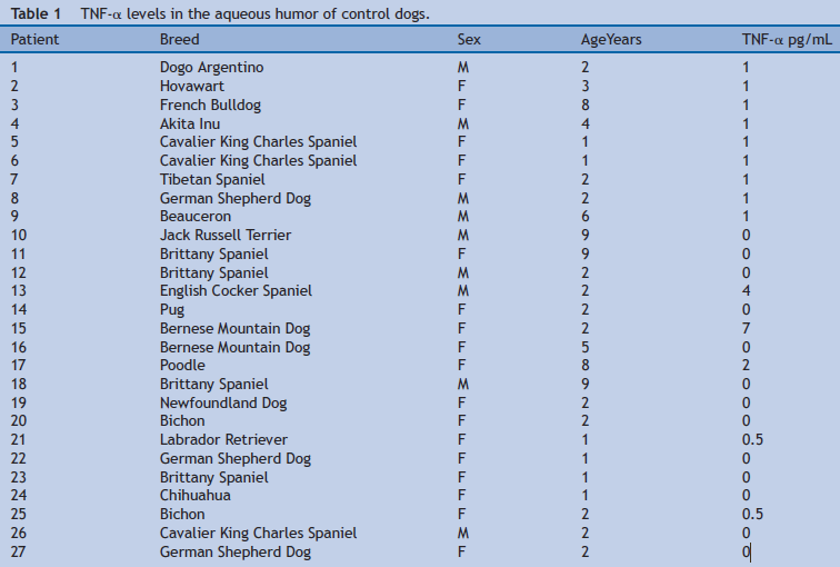
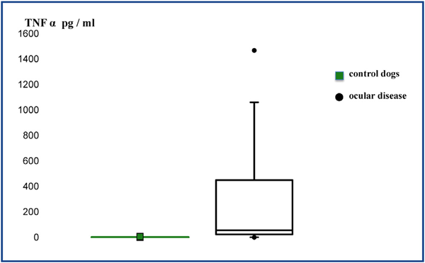
Figure 1 Box plot diagram shows the concentration of TNF-α in the aqueous humor between the two groups.
 
figure 2
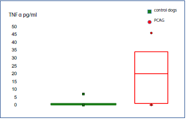
Figure 2 Box plot diagram shows the concentration of TNF-α in the aqueous humor of dogs from the control group and with primary angle closure glaucoma (PCAG).

TNF-α levels were significantly higher (P = 0.001) in the aqueous humor of dogs from the group with AAU (mean = 511.3; median = 372; IQR = 584) compared to the control group (mean = 1.1; median = 0.5; IQR = 1) (Fig. 3).

figure 3
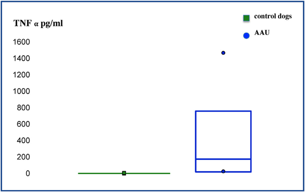
Figure 3 Box plot diagram shows the concentration of TNF-α in the aqueous humor of dogs from the control group and with acute anterior uveitis (AAU).

In the group with ocular disease, TNF-α concentrations (Table 2) were compared between the group of dogs with PCAG (mean = 19.4; median = 20; IQR = 33) and the group of dogs with AAU (mean = 511.3; median = 372; IQR = 584), TNF-α levels were higher in the aqueous humor of the uveitis group (P = 0.001) (Fig. 4).

table 2
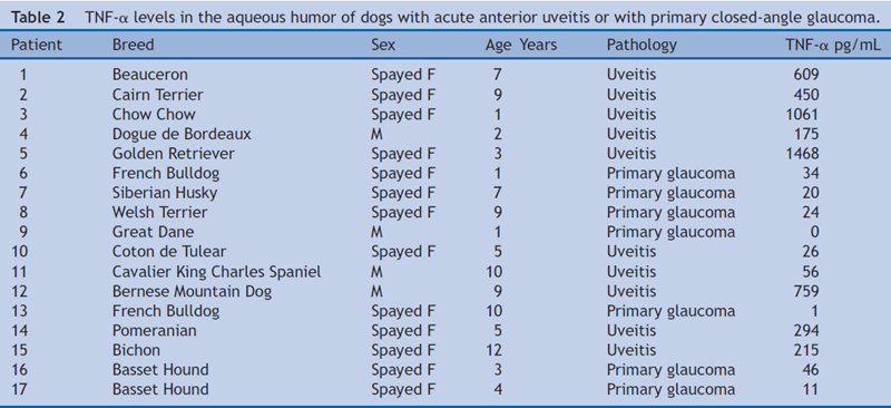
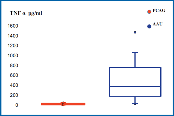
Figure 4 Box plot diagram shows the concentration of TNF-α in the aqueous humor of dogs with acute anterior uveitis (AAU) or primary closed-angle glaucoma (PCAG).

None of the dogs in the normal control group showed any ocular complications attributable to the anterior chamber oculocentesis on day 15 after sampling.

Discussion

Our study shows for the first time, to the authors’ knowledge, the feasibility of measuring TNF-α in the aqueous humor of dogs in private practice.

Our study also highlights the ease of removal of the aqueous humor and the perfect harmlessness of oculocentesis for the dog. No complication related to this act has been revealed among the group of the control dogs after eye examinations during the fifteen days following collection. In veterinary ophthalmology, as it was already the case in human ophthalmology, anterior chamber oculocentesis for diagnostic purposes becomes a recognised technique. There are numerous and diverse infectious, inflammatory, immune-mediated, neoplastic, and traumatic causes of anterior uveitis in dogs and extensive diagnostic testing is required to formulate treatment and prognostic advice relating to comfort and vision 2123.

As assumed the aqueous humor from dogs with AAU had significantly higher levels of TNF-α than control dogs (Fig. 3). Interestingly, we also found for the dogs with primary glaucoma higher levels of TNF-α than control dogs (Fig. 2).

An increased TNF-α is well-known in uveitis and has been demonstrated in humans and other animal models. Anti-TNF-α treatment became widely used for refractory non-infectious uveitis. On the contrary in the case of glaucoma that is much less clear. Thus, in a study in human, TNF-α could not be detected in the aqueous humor of 85% of glaucomatous eyes of the patients, which included patients with primary opened-angle glaucoma (POAG), with exfoliation glaucoma (ExG) and with normal tension glaucoma (NTG) 24. The authors did not find a relationship between the intraocular pressure and the TNF-α concentration. But in considering the glaucoma subtypes, TNF-α was detected in about 30% of patients with exfoliation glaucoma. The average intraocular pressure in the ExG group was the highest observed (25.8 ± 9.3 mmHg) among the others glaucoma subtypes. Intraocular stress, including high pressure and ischemia, could stimulate TNF-α production and could explain increased TNF-α levels, as it was the case for our dogs with PACG.

In dogs unlike humans, glaucoma are mostly closed-angle glaucoma. During decompensation, increased pressure is brutal with very high-pressure levels, as opposed to what happens in humans. Vision loss is rapid. This hereditary glaucoma is linked to goniodysplasia and reaches both eyes, although the pathological involvement is delayed in time. The pressure is very high and these eyes quickly become buphthalmic in chronic phase and medical treatment has little influence on the high pressures 25. TNF-α was detected in about 80% of the glaucomatous eyes and is significantly increased in the group of glaucomatous dogs but extremely high levels of intraocular pressure should alone be able to explain this rise. Tissular stress must be very important at this pressure level and not only at the level of ganglion cells of the retina.

The inclusion of glaucomatous dogs chronic stage in our study was intended only to compare the levels of TNF-α in aqueous humor with those of the control group dogs and those of the group of dogs with AAU. We were able to show that chronic glaucoma dogs were more likely to have detectable levels of TNF-α in their aqueous humor but further investigations could be made in the future.

The aqueous humor from all our dogs with AAU has significantly higher levels of TNF-α than control dogs (Fig. 3). As we said in the introduction, to the authors’ knowledge there is only one experimental study on the measurement of TNF-α in the aqueous humor of uveitic dogs 18. They used a similar Elisa kit. In this study, TNF-α concentration in aqueous humor was below the detection limit for all dogs excepting very low levels for some dogs. They used repeated oculocentesis and consequent collapse of the anterior chamber under general anaesthesia to induce experimental anterior uveitis without causing obvious signs of ocular pain. Examination of the eyes following paracentesis did not reveal any blepharospasm or protrusion of the third eyelid and the Hogan Kimura score was 0 19. All our dogs presenting with uveitis scored 4. The difference in the severity of the uveitis could explain the higher TNF-α levels in our results.

In the intraocular disease group, we also noted a significant higher increase in TNF-α concentration in the aqueous humor of dogs with AAU compared to dogs with PACG (Fig. 4). The more severe acute inflammation in dogs with AAU might explain the greater levels of TNF-α in their aqueous humor compared to the more chronic pathology of glaucomatous dogs. But a limitation of our study is that we wanted to compare two different pathological entities as their pathophysiology. Future studies with larger dog cohorts are necessary and need to take into account differences in aetiological classification and in the grade of the intraocular disease involved but also kinetic factors inherent in the evolution of the disease.

Conclusion

In conclusion, our results show the feasibility of measuring TNF-α concentration in the aqueous humor of dogs using an Elisa kit. Dogs seem to be good animal models for subsequent human trials in the field of intraocular inflammation using TNF-α as a biomarker. Disclosure of interest The authors declare that they have no conflict of interest concerning this article.

References

  1. Khera TK, Dick AD, Nicholson LB. Mechanisms of TNF-! regulation in uveitis: focus on RNA-binding proteins. Prog Retin Eye Res 2010;29:610—21.
  2. Idriss HT, Naismith JH. TNF-alpha and the TNF receptor superfamily: structure-function relationship(s). Microsc Res Tech 2000;50:184—95.
  3. Wang H, Czura CJ, Tracey KJ, Angus WT, Michael TL. Tumor necrosis factor. In: Thomson AW, Lotze MT, editors. The cytokine handbook. 4th ed. London: Academic Press; 2003. p. 837—60.
  4. Hehlgans T, Mannel DN. The TNF-TNF receptor system. J Biol Chem 2002;383:1581—5.
  5. Lacomba MS, Martin CM, Gallardo Galera JM, Gomez Vidal MA, Estevez EC, Chamond RR, et al. Aqueous humor and serum tumor necrosis factor-alpha in clinical uveitis. Ophthalmic Res 2011;33:251—5.
  6. Ahn JK, Yu HG, Chung H, Park YG. Intraocular cytokine environment in active Behcet uveitis. Am J Ophthalmol 2006;142:429—34.
  7. Zorena K, Mysliwska J, Mysliwiec M, Balcerska A, Hak L, Lipowski P, et al. Serum TNF-alpha level predicts nonproliferative diabetic retinopathy in children. Mediators Inflamm 2007;92196:1—5.
  8. Gupta SK, Agarwal R, Srivastava S, Agarwal P, Agrawal SS, Saxena R, et al. The anti-inflammatory effects of Curcuma longa and Berberis aristata in endotoxin-induced uveitis in rabbits. Invest Ophthalmol Vis Sci 2008;49:4036—40.
  9. Mackensen F, Lutz T. Therapy for childhood uveitis: biologics: too often—–too late? Der Ophthalmologe 2011;108:213—21.
  10. Tu KL, Kaye SB, Sidaras G, Taylor W, Shenkin A. Effect of intraocular surgery and ketamine on aqueous and serum cytokines. Mol Vis 2007;13:1130—7.
  11. Han L, Song E, Dong Y, Dong Q, Yu J. Clinical significance of detection of TNF-! and IL-8in rabbits after IOL implantation. Journal of Jilin University Medicine 2004;30:94—6.
  12. Cvenkel B, Kopitar AN, Ihan A. Inflammatory Molecules in aqueous humor and on ocular surface and glaucoma surgery outcome. Mediators Inflamm 2010;2010:939602 [sous presse].
  13. André C, Plassais J. Le chien dans la pathologie et la génétique comparée : exemples de maladies et de genes partagés entre l’homme et le chien. Bull Acad Vet France 2012;165: 215—24.
  14. Regnier A, Whitley RD, Benard P, Bonnefoi M. Effect of flunixin meglumine on the breakdown of the blood-aqueous barrier following paracentesis in the canine eye. J Ocul Pharmacol 1986;2:165—70.
  15. Krohne SD, Vestre WA. Effects of flunixin meglumine and dexamethasone on aqueous protein values after intraocular surgery in the dog. Am J Vet Res 1987;48:420—2.
  16. Gilmour MA, Lehenbauer TW. Comparison of tepoxalin, carprofen and meloxicam for reducing intraocular inflammation in dogs. Am J Vet Res 2009;70:902—7.
  17. Dziezyc J, Millichamp NJ, Rohde BH, Smithy WB. Comparison of prednisolone and RMI-1068 in the ocular irritative response in dogs. Invest Ophthalmol Vis Sci 1992;33:460—5.
  18. Pinard CL, Gauvin D, Moreau M, Martel-Pelletier J, Pelletier JP, Troncy E. Measurements of canine aqueous humor inflammatory mediators and the effect of carprofen following anterior chamber paracentesis. Vet Ophthalmol 2011;14:296—303.
  19. Hogan MJ, Kimura SJ, Thygeson P. Signs and symptoms of uveitis. I. Anterior uveitis. Am J Ophthalmol 1959;47:155—70.
  20. Zapryanova D, Mircheva T, Denev SA. Plasma protein profiles and fibrinogen concentrations in dogs with experimentally induced Staphylococcus aureus infection. Rev Med Vet 2013;164:150—5.
  21. Van der Lelij A, Rothova A. Diagnostic anterior chamber paracentesis in uveitis: a safe procedure? Br J Ophthalmol 1997;81:976—9.
  22. Sandberg CA, Herring IP, Huckle WR, LeRoith T, Pickett JP, Rossmeisl JH. Aqueous humor vascular endothelial growth factor in dogs: association with intraocular disease and the development of pre-iridal fibrovascular membrane. Vet Ophthalmol 2012;15:21—30.
  23. Wiggans KT, Vernau W, Lappin MR, Thomasy SM, Maggs DJ. Diagnostic utility of Aqueocentesis and aqueous humor analysis in dogs and cats anterior uveitis. Vet Ophthalmol 2013, http://dx.doi.org/10.1111/vop.12075 [sous presse].
  24. Sawada H, Fukuchi T, Tanaka T, Abe H. Tumor necrosis factoralpha concentrations in the aqueous humor of patients with glaucoma. Invest Ophthalmol Vis Sci 2010;51:903—6.
  25. Plummer CE, Regnier A, Gelatt KN. The canine glaucomas. In: Gelatt KN, Gilger BC, Kern TJ, editors. Veterinary ophthalmology. 5th ed. Ames: John Wiley & Sons, Inc.; 2013. p. 1074—80.
Retour

Informations pratiques

CHV des Cordeliers

Retour
  • Lun
    8h - 12h 13h30 - 19h
  • Mar
    8h - 12h 13h30 - 19h
  • Merc
    8h - 12h 13h30 - 19h
  • Jeu
    8h - 12h 13h30 - 19h
  • Vend
    8h - 12h 13h30 - 19h
  • Sam
    8h - 12h 13h30 - 17h30
  • Dim
    Urgences 24h/7j
Retour

Adresse de la clinique

29 - 35 Av. du Maréchal Joffre 77100 MEAUX
retrouvez-nous sur Google Maps